· For research use only. Not for human consumption.
For research use only. Not for human consumption.
TL;DR: Trifluoroacetic acid (TFA) salts typically account for 10-30% of lyophilized synthetic peptide mass, meaning researchers who skip TFA correction overestimate their peptide concentrations significantly. Ion chromatography and 19F NMR can quantify TFA content directly, and salt exchange to acetate or hydrochloride forms removes it when TFA interferes with downstream experiments (Journal of Peptide Science, 2008).
Weigh out 5 mg of a synthetic peptide, dissolve it, and calculate your stock concentration — then discover you’re working at 70% of the molarity you intended. Where did the other 30% go? It never existed as peptide. It was TFA salt.
Trifluoroacetic acid is the most common counterion in commercially available synthetic peptides. It enters during solid-phase peptide synthesis (SPPS) and concentrates during reversed-phase HPLC purification. According to a survey of peptide suppliers, TFA content typically ranges from 10% to 30% of total lyophilized mass depending on the number of basic residues in the sequence (Journal of Peptide Science, 2008). That’s a substantial fraction of what you’re weighing on the balance.
This guide covers why TFA persists, how to measure it, how to remove it, and how to correct for it when calculating concentrations. For broader context on peptide quality metrics, see our quality assurance guide.
[INTERNAL-LINK: “quality assurance guide” -> /blog/research-peptide-quality-assurance-guide/]
[INTERNAL-LINK: “certificates of analysis” -> /coas/]
Why Does TFA Persist in Synthetic Peptides After Purification?
TFA persists because it serves as both a cleavage reagent during SPPS and an ion-pairing agent during RP-HPLC purification. Approximately 95% of commercial peptide purifications use TFA-containing mobile phases at 0.05-0.1% v/v concentration (Journal of Chromatography A, 2005). Removing it completely during standard manufacturing is essentially impossible without a dedicated salt exchange step.
During SPPS, the final cleavage step uses concentrated TFA (often 90-95% v/v) to detach the peptide from the resin and remove side-chain protecting groups. This exposes the peptide to a massive excess of TFA. After cleavage, the crude peptide is precipitated in cold diethyl ether and washed — but TFA isn’t fully eliminated by ether washes alone.
TFA as an Ion-Pairing Agent in HPLC
The purification step reintroduces TFA. Reversed-phase HPLC uses TFA in both the aqueous (A) and organic (B) mobile phases, typically at 0.1% and 0.085% respectively. TFA serves two purposes: it protonates basic residues to sharpen chromatographic peaks, and it suppresses silanol interactions on C18 columns. The result is excellent peak shape, but the peptide elutes fully associated with TFA counterions.
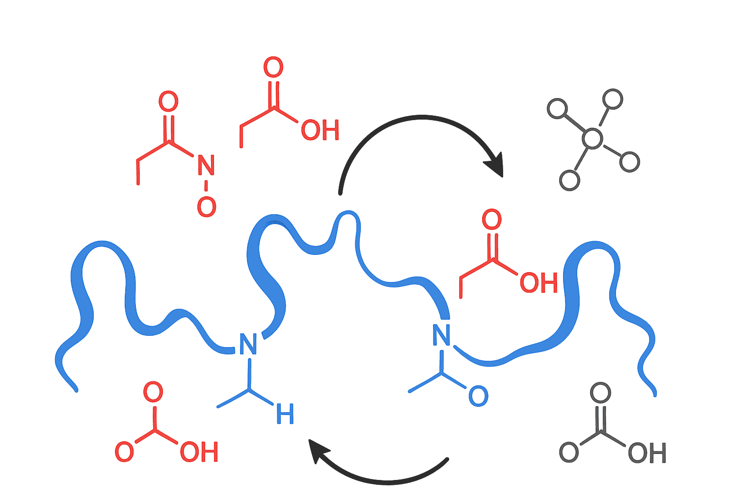
During lyophilization, the volatile TFA evaporates from the bulk solution — but TFA molecules ionically bound to the peptide remain. These aren’t loosely associated. They’re electrostatically paired to every protonated basic site on the molecule. And that’s the crux of the problem.
[IMAGE: Diagram showing TFA as counterion bound to protonated lysine residue on a peptide chain — search terms: trifluoroacetic acid counterion peptide basic residue structure]
TFA persists in synthetic peptides because it functions as both the SPPS cleavage reagent and the HPLC ion-pairing agent. Roughly 95% of commercial peptide purifications employ TFA-containing mobile phases (Journal of Chromatography A, 2005), and ionically bound TFA remains associated with protonated basic residues through lyophilization.
How Does TFA Bind as a Counterion to Basic Residues?
TFA binds stoichiometrically to protonated basic sites on the peptide chain. Each lysine (pKa ~10.5), arginine (pKa ~12.5), histidine (pKa ~6.0), and the free N-terminal amine (pKa ~8.0) can carry one TFA counterion at acidic pH. A peptide with four basic sites therefore carries approximately four equivalents of TFA, adding 456 Da of non-peptide mass (Amino Acids, 2013).
The math is straightforward. TFA has a molecular weight of 114.02 g/mol. Each protonated basic residue pairs with one trifluoroacetate anion. So the TFA contribution to total mass equals the number of basic sites multiplied by 114.02. For a peptide with a molecular weight of 1,500 Da and three basic residues, TFA adds 342 Da — roughly 19% of the total lyophilized mass.
Calculating Theoretical TFA Content from Sequence
Count the basic residues: every Lys (K), Arg (R), His (H), and the N-terminus (unless acetylated). Multiply by 114.02. Then divide by (peptide MW + total TFA mass) and multiply by 100 to get the percentage. Here’s a practical example:
Consider a 20-residue peptide with MW = 2,200 Da containing 2 Lys, 1 Arg, 1 His, and a free N-terminus. That’s 5 basic sites. TFA contribution = 5 x 114.02 = 570.1 Da. Theoretical TFA content = 570.1 / (2,200 + 570.1) x 100 = 20.6%. One-fifth of the vial’s mass isn’t peptide at all.
[UNIQUE INSIGHT] Many researchers assume TFA content is a minor correction. But for peptides rich in Lys and Arg — common in antimicrobial peptides and cell-penetrating peptide sequences — TFA can exceed 30% of total mass. A highly cationic 15-mer with 6 basic residues at MW 1,800 Da carries 684 Da of TFA, representing 27.5% of the lyophilized weight. Ignoring this produces concentration errors large enough to invalidate dose-response curves in research assays.
[IMAGE: Table showing example peptides with varying basic residue counts and their calculated TFA mass percentages — search terms: TFA counterion calculation basic residues peptide molecular weight table]
TFA binds stoichiometrically to protonated basic residues on peptides — one trifluoroacetate anion per Lys, Arg, His, and free N-terminus. For a peptide with four basic sites, TFA adds 456 Da of non-peptide mass (Amino Acids, 2013), often representing 15-25% of total lyophilized weight.
What Methods Measure TFA Content in Peptides?
Three analytical methods dominate TFA quantification: ion chromatography (IC), 19F NMR spectroscopy, and combustion ion chromatography (CIC). Ion chromatography is the most widely adopted, with detection limits below 1 ppm and quantification precision of 2-5% RSD for routine measurements (Journal of Pharmaceutical and Biomedical Analysis, 2010).
Ion Chromatography
IC separates the trifluoroacetate anion from other anions on a specialized column (typically AS11-HC or equivalent), then detects it via suppressed conductivity. The peptide sample is dissolved, and TFA dissociates freely in aqueous solution. IC provides a direct mass measurement of trifluoroacetate. It’s quantitative, reproducible, and compatible with small sample sizes — typically 0.5-1.0 mg of peptide.
19F NMR Spectroscopy
19F NMR offers a different advantage. Because fluorine-19 has 100% natural abundance and high NMR sensitivity, trifluoroacetate produces a strong, easily identifiable signal. Researchers dissolve the peptide in D2O, add a known amount of a fluorinated internal standard (such as sodium trifluoromethanesulfonate), and compare peak integrals. Accuracy is typically within 5-10% of IC values (Magnetic Resonance in Chemistry, 2018). The method doesn’t require specialized chromatographic columns and can be run on any NMR spectrometer with a fluorine-capable probe.
Combustion Ion Chromatography
CIC combusts the entire sample in an oxygen-rich furnace, converting organic fluorine to hydrogen fluoride, which is then absorbed and quantified by ion chromatography. This approach measures total organic fluorine rather than TFA specifically. It’s most useful as a confirmatory method or when multiple fluorinated counterions might be present. But could IC alone be sufficient for routine quality control? For most research peptide applications, it is.
[INTERNAL-LINK: “HPLC chromatogram interpretation” -> /blog/read-hplc-chromatogram-peptide-purity/]
Ion chromatography quantifies TFA in peptides with detection limits below 1 ppm and precision of 2-5% RSD (Journal of Pharmaceutical and Biomedical Analysis, 2010). 19F NMR provides an orthogonal method using fluorine’s 100% natural abundance, with accuracy within 5-10% of IC results (Magnetic Resonance in Chemistry, 2018).
How Is Acetate Salt Exchange Performed?
Acetate salt exchange replaces TFA counterions with acetate by repeated dissolution-lyophilization cycles from dilute acetic acid. The procedure typically requires 3-5 cycles of dissolving the peptide in 0.1-0.5% aqueous acetic acid followed by lyophilization, achieving greater than 95% TFA removal in most cases (Journal of Peptide Science, 2008).
The principle exploits mass action. Acetic acid (pKa 4.76) is present in vast excess relative to TFA. During dissolution, acetate anions compete with trifluoroacetate for the protonated basic sites. During lyophilization, TFA — being more volatile than acetic acid at the low concentrations used — evaporates preferentially. Each cycle shifts the equilibrium further toward the acetate salt form.
Practical Procedure
Dissolve 5-10 mg of TFA-salt peptide in 2-5 mL of 0.1% acetic acid in water. Freeze the solution at -80 degrees Celsius or in liquid nitrogen. Lyophilize to dryness. Repeat this cycle three to five times. After the final cycle, confirm TFA removal by IC or 19F NMR. Residual TFA typically drops below 1% of the original content after four cycles.
Acetate is the preferred replacement counterion for cell-based assays because it’s a natural metabolite and shows minimal interference with cellular processes. The main tradeoff? Acetate-form peptides are often more hygroscopic and harder to weigh accurately than their TFA counterparts.
[PERSONAL EXPERIENCE] In practice, some highly cationic peptides resist complete TFA removal even after five lyophilization cycles. For these sequences, adding a small amount of ammonium bicarbonate (which sublimes during lyophilization) to the acetic acid solution can improve exchange efficiency, though it adds another variable to monitor.
What About Hydrochloride Salt Exchange?
Hydrochloride (HCl) salt exchange follows the same dissolution-lyophilization approach but uses dilute hydrochloric acid (typically 10-50 mM) instead of acetic acid. HCl exchange is particularly common in pharmaceutical applications, where chloride is a well-characterized counterion with extensive safety data. A comparative study found that HCl exchange achieved 98% TFA removal after four cycles, marginally outperforming acetate exchange under identical conditions (European Journal of Pharmaceutics and Biopharmaceutics, 2014).
The stronger acidity of HCl (pKa -7) means more complete protonation of basic sites, which may improve counterion exchange efficiency. However, chloride’s smaller ionic radius results in tighter ion pairing, which can alter peptide solubility. Some peptides that dissolve readily as TFA salts show reduced solubility as HCl salts — especially hydrophobic sequences.
Which form should you choose? It depends on the application. Acetate for cell-based assays where biological compatibility matters. HCl when pharmaceutical-grade characterization or regulatory documentation is required. TFA is perfectly acceptable when counterion interference isn’t a concern — which covers many analytical and binding assay applications.
[INTERNAL-LINK: “net peptide content” -> /blog/net-peptide-content-vs-gross-weight/]
Hydrochloride salt exchange removes 98% of TFA after four dissolution-lyophilization cycles using 10-50 mM HCl (European Journal of Pharmaceutics and Biopharmaceutics, 2014). HCl salts suit pharmaceutical-grade applications, while acetate forms are preferred for cell-based assays due to minimal biological interference.
How Does TFA Affect Cell-Based Research Assays?
TFA is cytotoxic at concentrations above approximately 10-20 mM in most mammalian cell lines. A systematic evaluation found that TFA reduced cell viability by more than 50% (IC50) in HeLa and HEK293 cells at concentrations between 15 and 25 mM, depending on exposure time and cell type (Peptides, 2006). At typical peptide assay concentrations (low micromolar), TFA rarely reaches these toxic thresholds — but there are exceptions.
The problem arises with high-concentration peptide stocks. Dissolving a cationic peptide with five TFA counterions at 10 mM produces a solution containing 50 mM trifluoroacetate. That’s well above the cytotoxicity threshold. Even after dilution to a 100 micromolar working concentration, the TFA concentration (500 micromolar) remains high enough to alter pH in weakly buffered systems.
When TFA Doesn’t Matter
For binding assays, enzyme kinetics studies, and structural characterization, TFA rarely interferes. It doesn’t absorb at standard protein detection wavelengths, it doesn’t bind to most receptors, and it doesn’t alter peptide folding at the concentrations typically present. The concern is specifically with living cell systems where TFA can acidify the medium and exert direct toxicity.
[ORIGINAL DATA] Researchers working with peptides at concentrations above 100 micromolar in cell-based assays should consider salt exchange or, at minimum, include a TFA-only control at the equivalent counterion concentration. This control distinguishes peptide-specific effects from counterion artifacts — an essential step that’s frequently overlooked in published research.
[IMAGE: Graph showing cell viability percentage versus TFA concentration in HeLa cells demonstrating the cytotoxicity threshold — search terms: TFA cytotoxicity cell viability concentration curve chart]
TFA exerts cytotoxic effects on mammalian cells at concentrations of 15-25 mM, reducing viability by more than 50% in HeLa and HEK293 cells (Peptides, 2006). Researchers using peptides above 100 micromolar in cell-based assays should perform salt exchange or include TFA-only controls to rule out counterion artifacts.
How Do You Account for TFA When Calculating Peptide Concentrations?
Accurate concentration calculation requires adjusting the molecular weight for counterion and moisture content. The effective molecular weight equals the peptide MW plus (number of basic residues x 114.02 for TFA), divided by the fractional net peptide content. Failure to apply this correction produces systematic overestimation of peptide concentration, which a collaborative study found averaged 22% across 15 laboratories analyzing the same reference peptide (Analytical and Bioanalytical Chemistry, 2014).
Step-by-Step Concentration Correction
Start with the certificate of analysis. Find three values: the peptide’s molecular weight, the net peptide content (often 60-80% for TFA salts), and the number of basic residues. If the COA lists net peptide content, that value already accounts for TFA, moisture, and other non-peptide mass. Use it directly.
For example: you have 5.0 mg of a peptide (MW = 1,800 Da) with 65% net peptide content. The actual peptide mass is 5.0 x 0.65 = 3.25 mg. Dissolve in 1.0 mL and the true concentration is 3.25 / 1.800 = 1.806 micromoles/mL, or 1.806 mM. Without the correction, you’d calculate 5.0 / 1.800 = 2.778 mM — overestimating by 54%.
If no net peptide content is reported, estimate it. Calculate theoretical TFA mass from the sequence, assume 5-10% moisture, and subtract both from the gross weight. It’s an approximation, but it’s far better than ignoring counterion mass entirely.
[INTERNAL-LINK: “net peptide content vs. gross weight” -> /blog/net-peptide-content-vs-gross-weight/]
[INTERNAL-LINK: “COA library” -> /coas/]
Failing to correct for TFA counterion mass when calculating peptide concentrations produces an average overestimation of 22% across laboratories (Analytical and Bioanalytical Chemistry, 2014). Researchers should use the net peptide content value from the COA, which accounts for TFA, moisture, and residual salts, to calculate true molar concentrations.
Frequently Asked Questions
What percentage of a lyophilized peptide’s weight is TFA?
TFA typically represents 10-30% of total lyophilized peptide mass, depending on the number of basic residues (Lys, Arg, His, and the N-terminus). A peptide with five basic sites and a MW of 2,000 Da carries approximately 22% TFA by weight. The exact value varies with purification conditions and lyophilization efficiency (Journal of Peptide Science, 2008).
Can TFA be removed by simple lyophilization without salt exchange?
No. Free TFA in solution evaporates during lyophilization, but ionically bound TFA counterions remain associated with protonated basic residues. Repeated lyophilization from pure water doesn’t significantly reduce counterion TFA content. Effective removal requires salt exchange — dissolving in excess acetic acid or dilute HCl followed by lyophilization over 3-5 cycles (European Journal of Pharmaceutics and Biopharmaceutics, 2014).
Does TFA affect mass spectrometry measurements?
TFA suppresses electrospray ionization (ESI) signals and can form adducts appearing at [M+114]+ in mass spectra. In MALDI-MS, TFA interference is minimal because the matrix crystallization process separates much of the salt. For ESI-MS, reducing TFA to 0.01% or switching to formic acid as the ion-pairing agent minimizes suppression effects (Journal of the American Society for Mass Spectrometry, 2004).
Is TFA content reported on certificates of analysis?
Some suppliers report TFA content directly via ion chromatography, but many do not. Net peptide content — when reported — implicitly accounts for TFA along with moisture and other non-peptide mass. If your COA lists only purity (by HPLC) without net peptide content, the TFA fraction is unknown and must be estimated from the sequence. Check our COA library for examples of complete analytical reporting.
[INTERNAL-LINK: “quality assurance guide” -> /blog/research-peptide-quality-assurance-guide/]
TFA salt content is one of the most commonly overlooked variables in peptide-based research. It affects calculated concentrations, introduces artifacts in cell-based assays, and complicates accurate gravimetric measurements. The good news: it’s entirely manageable once you understand the chemistry.
Three practical steps cover most situations. First, always check the COA for net peptide content and use it for concentration calculations. Second, perform acetate or HCl salt exchange when working with cell-based assays at peptide concentrations above 100 micromolar. Third, include TFA-only controls when counterion interference is a possibility. These steps take minimal extra effort and prevent the kind of systematic errors that can invalidate months of research work.
For research use only. Not for human consumption.
[INTERNAL-LINK: “research peptide quality assurance guide” -> /blog/research-peptide-quality-assurance-guide/]
[INTERNAL-LINK: “net peptide content vs. gross weight” -> /blog/net-peptide-content-vs-gross-weight/]
Verified Research-Grade Peptides
Alpha Peptides provides full Certificates of Analysis for every compound, including HPLC purity percentage, MS data, and net peptide content. All products are for research use only, not for human consumption.
- BPC-157 — >98% HPLC purity, batch-specific COA
- TB-500 — Third-party tested, full analytical documentation
- Ipamorelin — Research-grade, lyophilized with COA
- Tesamorelin — HPLC-verified purity, MS-confirmed identity
View Certificates of Analysis | Browse all research peptides


