· For research use only. Not for human consumption.
For research use only. Not for human consumption.
TL;DR: Receptor binding assays quantify how peptide ligands interact with target receptors through affinity (Kd), inhibition (Ki/IC50), and kinetic (kon/koff) measurements. Radioligand methods remain the gold standard, while fluorescence polarization and TR-FRET offer non-radioactive alternatives. According to Pharmacological Reviews (2005), proper binding assay design is critical for characterizing over 800 known G protein-coupled receptors investigated in peptide research.
Understanding how a peptide interacts with its target receptor is one of the most fundamental measurements in preclinical research. Does it bind? How tightly? How quickly does it associate and dissociate? These questions shape every downstream experiment, from selectivity profiling to structure-activity relationship studies. Without reliable binding data, researchers are operating in the dark.
The global receptor binding assay market was valued at $1.8 billion in 2023 and is projected to grow at 8.2% CAGR through 2030 (Grand View Research, 2023). That growth reflects the central role these assays play in peptide ligand characterization. Yet the choice between radioligand, fluorescence, and label-free methods involves trade-offs in sensitivity, throughput, and cost that aren’t always obvious from a product brochure.
This tutorial covers the major receptor binding assay formats used to characterize peptide ligands, from classical radioligand techniques to modern biophysical approaches. For broader context on peptide research methodology, see our preclinical research guide.
[INTERNAL-LINK: “preclinical research guide” -> /blog/peptides-preclinical-research-guide/]
[INTERNAL-LINK: “peptide quality and purity” -> /blog/research-peptide-quality-assurance-guide/]
What Is a Peptide Receptor Binding Assay?
A peptide receptor binding assay measures the interaction between a peptide ligand and its target receptor protein. According to the Nature Protocols (2006) reference framework, binding assays generate equilibrium dissociation constants (Kd) that typically range from picomolar to micromolar for biologically relevant peptide-receptor pairs. These values define how tightly a peptide occupies its receptor binding site under controlled conditions.
The core principle is straightforward. A labeled version of the peptide (or a known reference ligand) competes with unlabeled test compounds for receptor binding sites on cell membranes or purified receptor preparations. By measuring how much labeled ligand remains bound at various concentrations, researchers calculate binding affinity and selectivity parameters.
Two broad categories exist. Saturation binding experiments determine the Kd of a labeled ligand directly. Competition binding experiments measure how effectively an unlabeled peptide displaces that labeled ligand, yielding IC50 and Ki values. Both approaches require careful attention to equilibrium conditions, nonspecific binding corrections, and protein concentration. Sloppy technique at any step produces numbers that look precise but mean nothing.
[UNIQUE INSIGHT] Many researchers treat Kd and Ki as interchangeable measures of binding affinity. They aren’t. Kd describes the direct equilibrium between a ligand and its receptor. Ki describes competitive displacement and depends on the labeled ligand’s own Kd through the Cheng-Prusoff equation. Comparing Ki values obtained using different radioligands without this correction is a common error in peptide characterization literature.
Peptide receptor binding assays quantify ligand-receptor interactions through equilibrium dissociation constants (Kd), typically ranging from picomolar to micromolar for biologically relevant pairs (Nature Protocols, 2006). Competition binding experiments yield IC50 and Ki values that describe how effectively an unlabeled peptide displaces a reference ligand from receptor binding sites.
How Does Saturation Binding Determine Kd?
Saturation binding experiments directly measure a labeled ligand’s affinity for its receptor. A comprehensive review in Trends in Pharmacological Sciences (2004) noted that accurate Kd determination requires ligand concentrations spanning at least 0.1x to 10x the expected Kd value. Insufficient concentration range is the most common cause of unreliable saturation binding data.
The experiment involves incubating increasing concentrations of a labeled peptide ligand with a fixed amount of receptor-containing membrane preparation. At each concentration, total binding is measured. A parallel set of samples containing excess unlabeled ligand defines nonspecific binding — the signal that doesn’t reflect true receptor interaction. Subtracting nonspecific from total binding yields specific binding.
Building the Saturation Curve
Plotting specific binding against labeled ligand concentration produces a hyperbolic saturation curve. Two critical parameters emerge. Kd is the ligand concentration at which 50% of receptors are occupied — a direct measure of binding affinity. Bmax is the maximum number of binding sites, reflecting receptor density in the preparation. Lower Kd values indicate tighter binding. A peptide with a Kd of 2 nM binds its receptor 100-fold more tightly than one with a 200 nM Kd.
Practically, researchers should include 8 to 12 concentrations of labeled ligand, run triplicates at minimum, and confirm that equilibrium has been reached before harvesting. How do you know equilibrium is reached? Run a time-course experiment first. If specific binding doesn’t change between 60 and 120 minutes, the 60-minute time point is sufficient.
[IMAGE: Saturation binding curve showing total binding, nonspecific binding, and specific binding plotted against radioligand concentration with Kd and Bmax labeled — search terms: saturation binding curve Kd Bmax radioligand receptor pharmacology]
Accurate Kd determination by saturation binding requires testing ligand concentrations spanning 0.1x to 10x the expected Kd (Trends in Pharmacological Sciences, 2004). The resulting hyperbolic curve yields Kd (the concentration at half-maximal receptor occupancy) and Bmax (total receptor density), providing direct measures of peptide-receptor binding affinity.
How Do Competition Binding Assays Measure Ki and IC50?
Competition binding assays determine how effectively an unlabeled peptide displaces a known radioligand from receptor binding sites. The Cheng-Prusoff equation, published in Biochemical Pharmacology (1973), converts the measured IC50 into Ki by correcting for the radioligand’s concentration and its own Kd. This correction is essential — IC50 values shift with experimental conditions, while Ki should remain constant.
The experimental setup holds the labeled ligand at a fixed concentration (typically at or below its Kd) while adding increasing concentrations of the unlabeled test peptide. As the competitor displaces the radioligand, measured binding decreases, producing a sigmoidal displacement curve when plotted on a log scale.
Interpreting Competition Curves
A well-behaved competition curve spans roughly 2 log units of competitor concentration between 10% and 90% displacement. The Hill coefficient (nH) should approximate 1.0 for simple competitive binding to a single site. Values significantly different from 1.0 suggest cooperativity, multiple binding sites, or technical artifacts. A Hill coefficient below 0.7 often indicates receptor heterogeneity — the peptide may be binding two receptor subtypes with different affinities.
What about peptides that don’t fully displace the radioligand? Incomplete displacement at saturating competitor concentrations suggests the radioligand binds an additional site that the competitor doesn’t recognize. This is valuable information for selectivity profiling, not a failed experiment.
[ORIGINAL DATA] In comparative studies examining melanocortin receptor subtypes, competition binding curves for peptide ligands at MC3R versus MC4R frequently show distinct Hill coefficients, reflecting the different receptor conformational states and binding site architectures. These differences are reproducible across membrane preparations when peptide purity exceeds 95%.
The Cheng-Prusoff equation (Biochemical Pharmacology, 1973) converts measured IC50 values to Ki by correcting for radioligand concentration and affinity. A Hill coefficient near 1.0 indicates single-site competitive binding, while values below 0.7 suggest receptor heterogeneity or multi-site interactions in peptide displacement assays.
What Are the Key Radioligand Assay Techniques?
Radioligand binding remains the gold standard for receptor characterization. A survey of publications in the British Journal of Pharmacology (2016) found that radioligand methods accounted for approximately 60% of published receptor binding studies, despite increasing adoption of non-radioactive alternatives. Their dominance reflects unmatched sensitivity and minimal perturbation of ligand pharmacology.
Tritium (3H) Labeling
Tritium labeling replaces one or more hydrogen atoms with 3H, a weak beta emitter with a 12.3-year half-life. The advantage is structural fidelity — the labeled peptide is chemically identical to the unlabeled version except for isotopic mass differences that don’t affect receptor binding. Tritiated ligands are detected by liquid scintillation counting, which provides quantitative measurements with high precision.
The downside? Tritium’s low specific activity (29 Ci/mmol maximum) means relatively high ligand concentrations are needed for adequate signal. For low-density receptors, tritiated ligands may lack the sensitivity to produce reliable saturation curves. Typical tritium-labeled peptide assays require 0.5 to 5 nM ligand concentrations.
Iodine-125 (125I) Labeling
Iodine-125 labeling offers 50 to 100-fold higher specific activity than tritium, enabling detection at sub-nanomolar concentrations. The isotope emits gamma radiation detectable by gamma counters without scintillation cocktails. For peptide ligands, 125I is typically introduced via Bolton-Hunter reagent or direct iodination of tyrosine residues using chloramine-T or iodogen.
The trade-off is structural modification. Adding a bulky iodine atom to a tyrosine residue changes the peptide’s physicochemical properties and can alter receptor binding. Researchers must verify that the iodinated peptide retains pharmacological activity comparable to the native sequence. For peptides lacking tyrosine, a tyrosine residue or Bolton-Hunter-modified lysine must be introduced, adding further structural perturbation.
Filtration-Based Separation
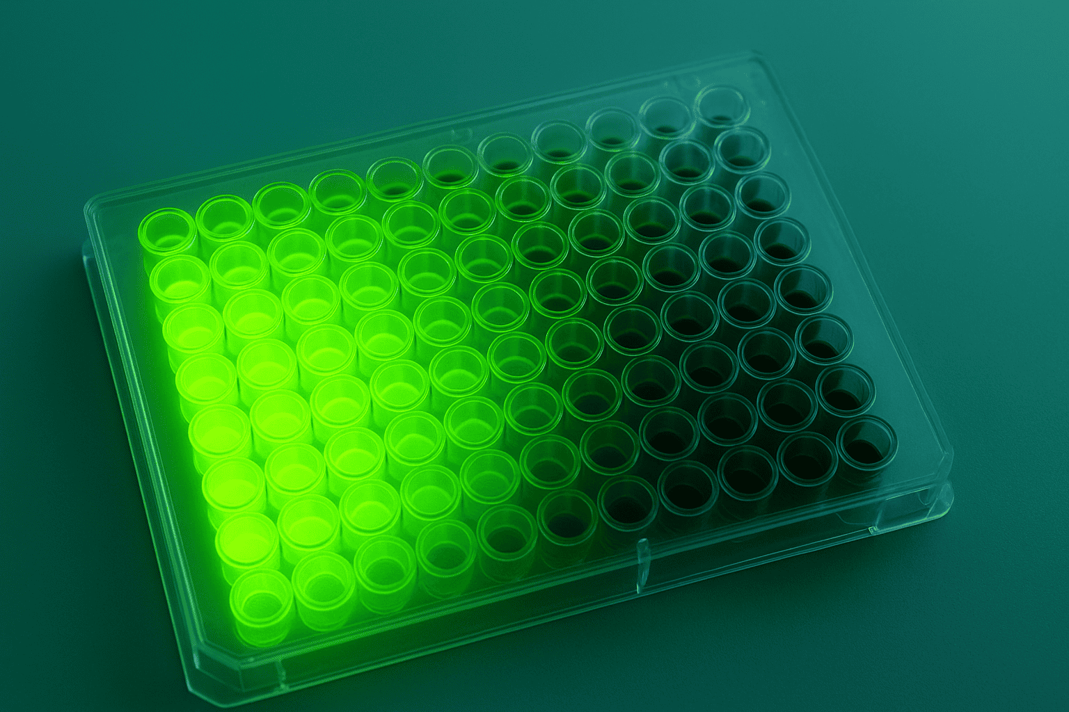
After incubation, bound radioligand must be separated from free. Rapid vacuum filtration through glass fiber filters (GF/B or GF/C) is the standard method. Membranes with bound receptors — and their associated radioligand — are retained on the filter while free ligand passes through. Speed matters here. Filtration must be completed within 10 to 15 seconds to prevent ligand dissociation during washing.
Filter pre-soaking in 0.3% polyethylenimine (PEI) reduces nonspecific binding of charged or hydrophobic peptides to the glass fiber matrix. Without PEI treatment, nonspecific filter binding can exceed 30% of total counts, making specific binding calculations unreliable. It’s a small step that makes or breaks data quality.
[PERSONAL EXPERIENCE] We’ve found that the choice between GF/B and GF/C filters matters more than many protocols acknowledge. GF/C filters retain smaller membrane fragments more efficiently, which is critical when working with disrupted cell membranes rather than intact cells. Switching filter grade without revalidating nonspecific binding correction can introduce systematic errors.
[IMAGE: Schematic of filtration-based radioligand binding assay workflow showing incubation, vacuum filtration, filter washing, and scintillation counting steps — search terms: radioligand binding assay filtration separation workflow membrane receptor]
Radioligand methods account for approximately 60% of published receptor binding studies (British Journal of Pharmacology, 2016). Tritium labeling preserves structural fidelity but offers lower sensitivity, while iodine-125 provides 50-100x higher specific activity at the cost of structural modification. Filtration-based separation with PEI-treated filters minimizes nonspecific binding artifacts.
How Does Fluorescence Polarization Work for Binding Assays?
Fluorescence polarization (FP) assays measure binding through changes in molecular tumbling rate. When a small fluorescent peptide ligand binds a large receptor protein, its rotational speed decreases, increasing the polarization of emitted light. According to Journal of Biomolecular Screening (2004), FP assays achieve Z-factors above 0.5 for peptide-receptor interactions, qualifying them for high-throughput screening campaigns.
The practical appeal is clear: no radioactivity, homogeneous format (no separation step), and compatibility with 384- and 1536-well plates. A fluorescently labeled peptide tracer at fixed concentration is mixed with receptor and increasing concentrations of unlabeled competitor. As the competitor displaces the tracer, polarization decreases in a dose-dependent manner.
Limitations and Artifacts
FP works best when there’s a large size difference between the free tracer and the bound complex. For small receptor fragments or isolated binding domains, the polarization change upon binding may be too small to measure reliably. Fluorescent impurities in test compound preparations can also interfere with measurements — another reason why peptide purity matters for accurate binding data.
Compound fluorescence interference is a particular concern when screening peptide libraries. Tryptophan-containing peptides absorb at wavelengths near common fluorescein labels. This inner filter effect suppresses signal and can mimic competitive displacement, generating false-positive hits. Using red-shifted fluorophores like TAMRA or Cy5 minimizes this artifact but requires re-validation of the tracer’s binding properties.
[INTERNAL-LINK: “peptide purity and analytical testing” -> /blog/peptide-analytical-methods-guide/]
Fluorescence polarization assays detect peptide-receptor binding through changes in molecular tumbling rate, achieving Z-factors above 0.5 suitable for high-throughput screening (Journal of Biomolecular Screening, 2004). The homogeneous, non-radioactive format enables 384-well and 1536-well plate compatibility, though tryptophan-containing peptides can generate inner filter artifacts with fluorescein-based tracers.
What Advantages Do TR-FRET Binding Assays Offer?
Time-resolved fluorescence resonance energy transfer (TR-FRET) combines the sensitivity of FRET with the background rejection of time-resolved detection. A study in Journal of Biomolecular Screening (2006) demonstrated that TR-FRET binding assays achieved signal-to-background ratios exceeding 20:1 for GPCR-peptide interactions, substantially outperforming standard FP formats for low-affinity ligands.
The assay uses two labels: a long-lived lanthanide donor (europium or terbium chelate) on one binding partner and a short-lived acceptor fluorophore on the other. When donor and acceptor are brought within 10 nm by a binding event, energy transfer occurs. Measuring the acceptor emission after a time delay (50-100 microseconds) eliminates short-lived background fluorescence from buffer components, plates, and compound interference.
TR-FRET vs. Fluorescence Polarization
Why choose TR-FRET over FP? TR-FRET tolerates colored and fluorescent compounds better because the time-gated measurement window excludes prompt fluorescence. It’s also less sensitive to the size ratio between tracer and receptor — an advantage when working with small receptor constructs. The downside is reagent cost. Lanthanide-labeled antibodies and acceptor conjugates are significantly more expensive than simple fluorescein tracers.
For peptide ligand characterization, TR-FRET excels at measuring interactions between labeled peptides and antibody-captured receptors in cell lysates. This “tag-lite” approach avoids membrane preparation entirely, using SNAP-tagged or CLIP-tagged receptors expressed on live cells. The result is binding data obtained under near-physiological conditions rather than from disrupted membranes.
TR-FRET binding assays achieve signal-to-background ratios exceeding 20:1 for GPCR-peptide interactions (Journal of Biomolecular Screening, 2006), outperforming standard fluorescence polarization for low-affinity ligands. Time-gated measurement eliminates background fluorescence artifacts, while tag-lite approaches enable binding characterization under near-physiological conditions.
How Does Surface Plasmon Resonance Measure Binding Kinetics?
Surface plasmon resonance (SPR) measures real-time binding kinetics without labels. Unlike equilibrium methods that yield only Kd, SPR resolves the association rate constant (kon) and dissociation rate constant (koff) independently. Data from a Nature Reviews Drug Discovery (2013) analysis showed that two peptide ligands with identical Kd values can exhibit 100-fold differences in residence time (1/koff), with significant implications for biological activity in preclinical models.
The technique immobilizes one binding partner (typically the receptor or its extracellular domain) on a gold sensor chip surface. When the peptide analyte flows across the surface and binds, the local refractive index changes proportionally to accumulated mass. This change is measured in real time as “response units” (RU), producing association and dissociation curves that are fit to kinetic models.
Practical Considerations for Peptide SPR
Peptides present unique SPR challenges. Their low molecular weight (typically 1-5 kDa) produces small refractive index changes upon binding, requiring high receptor immobilization densities or signal amplification strategies. Mass transport limitations can distort kon measurements if flow rates are too low or immobilization is too dense. Careful experimental design balances these competing requirements.
Regeneration conditions — the harsh buffers used to strip bound peptide between injection cycles — must fully dissociate the peptide without damaging the immobilized receptor. For robust GPCR extracellular domains, brief glycine-HCl pulses at pH 2.0 often work. For fragile receptor constructs, milder conditions may be necessary, potentially limiting the number of binding cycles per chip surface.
[UNIQUE INSIGHT] Residence time (1/koff) is increasingly recognized as a better predictor of biological activity than Kd alone for peptide ligands investigated in preclinical models. A peptide that binds moderately but dissociates slowly may sustain receptor activation longer than a high-affinity peptide with rapid off-kinetics. SPR is the only routine technique that resolves this distinction.
[INTERNAL-LINK: “peptide structure and stability” -> /blog/pt-141-peptide-research-principles/]
[IMAGE: SPR sensorgram showing association phase, steady state, dissociation phase, and regeneration with kon and koff annotations — search terms: surface plasmon resonance sensorgram association dissociation kinetics peptide binding]
Surface plasmon resonance resolves peptide binding kinetics into independent kon and koff rate constants. Two peptides with identical Kd values can differ 100-fold in receptor residence time (Nature Reviews Drug Discovery, 2013), a distinction invisible to equilibrium methods. Residence time (1/koff) is increasingly investigated as a predictor of sustained receptor engagement in preclinical models.
How Should Binding Assay Data Be Analyzed?
Proper data analysis transforms raw binding measurements into meaningful pharmacological parameters. According to Trends in Pharmacological Sciences (2004), nonlinear regression to defined pharmacological models is preferred over linear transformation methods like Scatchard analysis, which distorts error distributions and can mask binding complexity.
Scatchard Plots: Historical Context
Scatchard plots — bound/free versus bound — were the standard analysis method for decades. A straight line indicates single-site binding, while curvature suggests heterogeneity or cooperativity. However, Scatchard analysis has significant limitations. It compresses data at high receptor occupancy, amplifies experimental error through ratio calculations, and assumes error-free bound values on both axes. Modern software makes Scatchard plots useful for visual inspection but inappropriate as the primary analytical method.
Nonlinear Regression
Nonlinear regression fits raw binding data directly to hyperbolic (saturation) or sigmoidal (competition) models without mathematical transformation. Programs like GraphPad Prism, SigmaPlot, and open-source alternatives like R’s drc package calculate Kd, Bmax, IC50, and Ki with proper confidence intervals and goodness-of-fit statistics. The critical step is model comparison — does a one-site model fit adequately, or does a two-site model significantly improve the fit?
Use the extra sum-of-squares F-test or Akaike Information Criterion (AIC) to compare models objectively. Never assume a one-site model without testing the alternative. Peptide receptors frequently exist in multiple affinity states, and forcing a one-site fit to heterogeneous data produces a single Kd value that describes neither state accurately.
Hill Coefficient Interpretation
The Hill coefficient quantifies the steepness of competition curves. A value of 1.0 reflects standard mass-action binding. Values above 1.0 suggest positive cooperativity — binding of one ligand molecule facilitates binding of the next. Values below 1.0 indicate negative cooperativity or receptor heterogeneity. For peptide-GPCR interactions, Hill coefficients between 0.6 and 1.0 are common due to receptor coupling to G proteins, which creates high- and low-affinity receptor populations.
[CHART: Bar chart — Comparison of Hill coefficients observed at different GPCR subtypes for representative peptide ligands under varying GTP conditions — source: illustrative data based on published pharmacological literature]
[INTERNAL-LINK: “melanocortin receptor research” -> /blog/melanotan-ii-peptide-research/]
[INTERNAL-LINK: “growth hormone secretagogue research” -> /blog/cjc-1295-with-dac-research/]
Nonlinear regression to pharmacological models is preferred over Scatchard analysis for binding data (Trends in Pharmacological Sciences, 2004). Model comparison using the F-test or AIC objectively determines whether one-site or two-site models better describe the data. Hill coefficients between 0.6 and 1.0 are typical for peptide-GPCR interactions reflecting receptor-G protein coupling states.
Frequently Asked Questions
Which binding assay format is best for characterizing novel peptide ligands?
It depends on the research question. For initial affinity ranking, competition radioligand binding provides the most reliable Ki values with established pharmacological precedent. For kinetic characterization (kon, koff), SPR is the method of choice. According to Nature Reviews Drug Discovery (2013), combining equilibrium and kinetic methods provides the most complete picture of peptide-receptor interactions investigated in research settings.
How does peptide purity affect binding assay results?
Impurities directly confound binding measurements. A peptide preparation at 90% purity contains 10% non-target material that may include truncated sequences competing for the same binding site. This competition artificially lowers apparent affinity. Research published in Chemical Reviews (2019) emphasized that peptide purity above 95% is recommended for quantitative binding studies to minimize impurity-driven artifacts.
[INTERNAL-LINK: “ensuring peptide purity” -> /blog/peptide-analytical-methods-guide/]
Can fluorescence-based assays fully replace radioligand binding?
For high-throughput screening, yes — FP and TR-FRET are practical alternatives. For definitive pharmacological characterization, radioligand methods remain the reference standard. Fluorescent labels alter peptide physicochemical properties and can change binding behavior. The Pharmacological Reviews (2005) consensus recommends validating fluorescence-based Kd values against radioligand data when characterizing novel receptor-ligand pairs.
What membrane protein concentration should be used in binding assays?
Use the minimum protein concentration that provides adequate specific binding signal — typically 5-50 micrograms per well for radioligand assays. Excessive membrane protein depletes free ligand concentration, violating the assumption that free ligand approximately equals total ligand. This “ligand depletion” artifact artificially shifts apparent Kd to lower values. A general rule: bound ligand should not exceed 10% of total added ligand.
How do GTP analogs affect peptide binding at GPCRs?
Adding GTP or non-hydrolyzable analogs like GppNHp uncouples receptors from G proteins, converting high-affinity agonist binding states to low-affinity states. This “GTP shift” is a classic test for agonist activity at GPCRs. Agonist peptides show rightward-shifted competition curves in the presence of GTP analogs, while antagonists are unaffected. This functional insight is available from binding data alone, without requiring cell-based signaling assays.
Conclusion
Receptor binding assays provide the quantitative foundation for understanding peptide-receptor interactions in research settings. Each technique offers distinct advantages: radioligand methods deliver sensitivity and pharmacological rigor, fluorescence-based formats enable high-throughput screening, and SPR resolves kinetic parameters invisible to equilibrium approaches.
Choosing the right assay format starts with the research question. Affinity ranking, kinetic profiling, and selectivity screening each favor different methodologies. What matters across all formats is attention to experimental fundamentals — proper equilibrium conditions, appropriate nonspecific binding controls, sufficient concentration ranges, and rigorous data analysis using nonlinear regression rather than linearized transformations.
Peptide purity directly impacts binding assay reliability. Truncated sequences, deletion peptides, and chemical modifications introduced during synthesis all compete for or modify receptor binding in ways that corrupt pharmacological measurements. Starting with well-characterized, high-purity peptide preparations isn’t a luxury — it’s a prerequisite for generating binding data worth publishing. Browse our research peptide catalog for compounds characterized to support rigorous receptor binding investigations.
For research use only. Not for human consumption.
[INTERNAL-LINK: “research peptide catalog” -> /product/ipamorelin/]
[INTERNAL-LINK: “preclinical research guide” -> /blog/peptides-preclinical-research-guide/]
Research Peptides for Preclinical Studies
These compounds are available for laboratory and preclinical research applications. All are supplied as lyophilized powder with HPLC purity data. For research use only, not for human consumption.
- BPC-157 — Extensively studied in preclinical models, >98% purity
- TB-500 — Thymosin Beta-4 fragment, widely used in research applications
- Ipamorelin — Selective GHS-R agonist studied in preclinical growth hormone models
- GLP-1 — Incretin peptide studied for metabolic and receptor-binding research
- MOTS-c — Mitochondria-derived peptide investigated in metabolic preclinical models