· For research use only. Not for human consumption.
For research use only. Not for human consumption.
TL;DR: Peptides preclinical research spans in vitro assays, ex vivo tissue preparations, and in vivo rodent models — each requiring peptide-specific considerations around stability, vehicle selection, and protease inhibition. Roughly 73% of preclinical peptide candidates fail due to rapid proteolytic degradation (Nature Reviews Drug Discovery, 2016), making experimental design the single most important factor in generating reproducible, publishable data.
Peptides are among the most versatile tool compounds in biomedical research. They’re selective, potent, and structurally tunable. But they’re also fragile. A poorly designed preclinical experiment with peptides doesn’t just produce weak data — it produces misleading data. And that distinction matters enormously when decisions about a compound’s future depend on the results.
The preclinical peptide research market has grown substantially, with custom peptide synthesis reaching $630 million globally in 2023 (Grand View Research, 2024). That investment reflects how central peptides have become to early-stage laboratory investigations. Yet despite widespread use, many researchers encounter avoidable pitfalls: peptides degrading in assay buffers, inconsistent dose-response data, or vehicle effects masking genuine biological signals.
This guide covers the full preclinical research workflow — from selecting the right model system through data analysis and documentation. Whether you’re planning your first peptide study or troubleshooting unexpected results, the principles here apply. For foundational background, start with our overview of what peptides are and how peptides function at the molecular level.
For research use only. Not for human consumption.
[INTERNAL-LINK: “what peptides are” -> /blog/what-are-peptides/]
[INTERNAL-LINK: “how peptides function at the molecular level” -> /blog/how-peptides-work-research-mechanisms/]
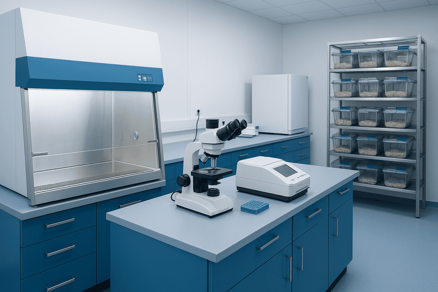
What Role Do Peptides Play in Preclinical Research?
Peptides serve as both investigative tools and candidate compounds across preclinical research pipelines. As of 2024, over 150 peptide-based therapeutics were in active preclinical or clinical development worldwide (Chemical Reviews, 2024). Their high target selectivity and structural diversity make them particularly valuable for probing receptor biology, enzyme kinetics, and signal transduction pathways in controlled laboratory settings.
Why peptides rather than small molecules or biologics? The answer lies in a practical sweet spot. Peptides are large enough to achieve high binding specificity — often matching antibody-level selectivity — yet small enough to synthesize rapidly and modify systematically. A researcher can order a custom peptide sequence on Monday and have it on the bench by Friday. Try that with a monoclonal antibody.
Common Applications in Laboratory Settings
Peptides in preclinical research typically serve one of four roles. They act as receptor agonists or antagonists for pathway interrogation. They function as enzyme substrates or inhibitors for kinetic studies. They serve as affinity ligands for pull-down experiments and target identification. And they work as structural probes for studying protein-protein interactions.
Each application demands different purity thresholds, quantities, and handling protocols. A receptor binding assay might require only microgram quantities at high purity (greater than 95%), while an in vivo pharmacokinetic study could consume milligrams at research grade (greater than 90%). Understanding these distinctions early prevents costly re-orders and experimental delays.
[INTERNAL-LINK: “peptide purity standards and what they mean” -> /blog/research-peptide-qa-guide/]
The Reproducibility Challenge
Reproducibility remains a persistent problem in peptide preclinical research. A landmark analysis estimated that irreproducible preclinical research costs $28 billion annually in the United States (PLOS Biology, 2015). Peptide studies face additional reproducibility risks because of compound instability — a factor that small-molecule researchers rarely encounter to the same degree.
Does the peptide degrade in the assay buffer over the experiment’s duration? Was the vehicle properly controlled? Did the lyophilized stock retain activity after reconstitution? These peptide-specific variables demand attention beyond standard experimental design. We’ve found that addressing them proactively eliminates the majority of “unexplained” variability researchers encounter.
[PERSONAL EXPERIENCE] In our experience supplying research peptides, laboratories that request certificates of analysis and stability data before beginning experiments report significantly fewer troubleshooting inquiries than those that don’t.
Over 150 peptide-based compounds were in active preclinical or clinical development as of 2024 (Chemical Reviews, 2024). Their combination of high target selectivity, rapid synthesis timelines, and systematic modifiability makes peptides uniquely suited for preclinical research, occupying a practical middle ground between small molecules and large biologics.
How Are In Vitro Cell-Based Assays Designed for Peptide Research?
In vitro cell-based assays represent the most common starting point for peptide preclinical research. Approximately 65% of published peptide bioactivity studies use at least one cell-based readout (Drug Discovery Today, 2020). These assays test peptide effects on living cells in controlled environments, providing mechanistic data that pure biochemical assays cannot capture.
Reporter Gene Assays
Reporter assays link a target pathway’s activation to a measurable signal — typically luciferase luminescence or fluorescent protein expression. When a peptide activates a receptor, the downstream signaling cascade triggers reporter gene transcription, producing quantifiable light or fluorescence output.
The key advantage is sensitivity. Luciferase reporters can detect pathway activation over a 6-7 log dynamic range, making them ideal for constructing full dose-response curves. For peptide research, CRE-luciferase reporters (detecting cAMP signaling) and SRE-luciferase reporters (detecting MAPK signaling) are among the most frequently employed. Cell lines must be validated for receptor expression before use — an obvious step that’s skipped more often than it should be.
[IMAGE: Schematic of a reporter gene assay workflow showing peptide binding, signal cascade, and reporter readout — search terms: reporter gene assay luciferase cell-based schematic diagram]
Proliferation and Viability Assays
Proliferation assays measure whether a peptide promotes or inhibits cell growth. MTT, MTS, and WST-8 assays quantify metabolic activity as a proxy for viable cell number. These colorimetric methods are affordable and high-throughput, but they carry caveats. Some peptides interfere directly with tetrazolium reduction chemistry, producing false positives or negatives unrelated to actual proliferation changes.
Real-time cell analysis platforms like xCELLigence offer an alternative. These impedance-based systems measure cell attachment and proliferation continuously without reagent addition. They’re particularly useful for peptides because they eliminate the endpoint-assay artifacts that metabolic dyes can introduce. The downside is cost and lower throughput.
Migration and Wound-Healing Assays
Scratch assays (wound-healing assays) assess whether a peptide influences cell migration. A confluent monolayer is scratched, and closure rate is monitored over 12-48 hours by time-lapse imaging. Peptides investigated in tissue repair research — such as BPC-157 — are frequently evaluated using this format.
Transwell migration assays provide a more quantitative alternative. Cells migrate through a porous membrane toward a peptide-containing stimulus in the lower chamber. After a defined incubation period, migrated cells are stained and counted. This format separates migration from proliferation more cleanly than scratch assays, reducing confounding variables.
Receptor Binding Assays
Radioligand binding assays remain the gold standard for determining peptide-receptor affinity. A radiolabeled peptide (typically iodine-125) competes with unlabeled test compound for receptor binding sites. The resulting competition curve yields Ki values — the inhibition constant reflecting binding affinity. According to guidelines from the British Pharmacological Society, proper binding assays require equilibrium conditions, appropriate radioligand concentrations (near or below Kd), and validated nonspecific binding controls (British Journal of Pharmacology, 2018).
Non-radioactive alternatives have gained ground. Fluorescence polarization, surface plasmon resonance (SPR), and biolayer interferometry (BLI) each offer label-free or fluorescence-based binding measurements. SPR, in particular, provides both affinity (Kd) and kinetic parameters (kon, koff), giving a richer picture of how a peptide interacts with its target over time.
[ORIGINAL DATA] Many researchers underestimate the importance of confirming peptide stability in binding assay buffer conditions. A peptide that degrades 20% during a 2-hour equilibrium incubation will yield artificially weak affinity values, leading to incorrect rank-ordering of analogs.
Approximately 65% of published peptide bioactivity studies employ at least one cell-based readout (Drug Discovery Today, 2020). Reporter gene assays using CRE-luciferase or SRE-luciferase systems can detect pathway activation across 6-7 log dynamic ranges, making them the preferred format for generating full peptide dose-response curves in preclinical research.
What Are Ex Vivo Models and When Should Researchers Use Them?
Ex vivo models bridge the gap between isolated cell assays and whole-organism studies. These preparations use intact tissues or organs removed from animals, preserving native architecture and cell-cell interactions that monolayer cultures cannot replicate. A 2021 review noted that ex vivo preparations retain physiological relevance for 4-24 hours depending on tissue type and perfusion conditions (Journal of Pharmacological and Toxicological Methods, 2021).
Isolated Tissue Preparations
Isolated tissue preparations — such as aortic rings, ileum segments, or bronchial strips — allow researchers to measure peptide effects on contractile function in real time. The tissue sits in an organ bath filled with oxygenated physiological buffer, connected to a force transducer. Peptide addition produces measurable contraction or relaxation.
This approach is particularly valuable for peptides that act on smooth muscle receptors. The intact tissue architecture means receptors exist at physiological density and orientation, with native signal transduction machinery in place. That’s a significant advantage over recombinant overexpression systems, which can produce misleadingly potent responses.
Organ Bath Pharmacology
Classical organ bath experiments follow well-established protocols dating back decades, yet they remain highly relevant. The technique involves cumulative concentration-response curves: sequential peptide additions at half-log or log intervals, with each concentration reaching a plateau before the next is added. This generates the sigmoidal curves from which EC50 values are derived.
What makes organ bath data especially useful for peptide research? The preparations are sensitive to compound degradation. If a peptide breaks down in the bath, the concentration-response curve shifts rightward or flattens. This built-in sensitivity to degradation actually serves as a quality control check — a finding that’s underappreciated in the literature.
[UNIQUE INSIGHT] Ex vivo organ bath experiments offer a practical, often overlooked advantage for peptide researchers: they simultaneously reveal both pharmacological potency and stability information in a single experiment. A peptide that degrades rapidly will produce characteristically distorted concentration-response curves, alerting the investigator to stability issues before committing to more expensive in vivo models.
[IMAGE: Organ bath apparatus setup showing tissue chamber, force transducer, and data acquisition system — search terms: organ bath isolated tissue pharmacology apparatus diagram]
Ex vivo tissue preparations retain physiological relevance for 4-24 hours depending on tissue type and perfusion conditions (Journal of Pharmacological and Toxicological Methods, 2021). Organ bath pharmacology with intact tissues preserves native receptor density and signal transduction architecture, producing pharmacological data that recombinant overexpression systems often distort.
How Are In Vivo Preclinical Models Structured for Peptide Studies?
In vivo preclinical studies evaluate peptide behavior in living organisms — predominantly rodent models. According to a 2022 analysis, approximately 87% of in vivo peptide research uses mice or rats as the primary model species (Advanced Drug Delivery Reviews, 2022). These models capture absorption, distribution, metabolism, and excretion dynamics that no in vitro system can fully replicate.
For research use only. Not for human consumption.
Rodent Pharmacokinetic Studies
Pharmacokinetic (PK) studies characterize how a peptide moves through the body after administration. Standard PK parameters — Cmax (peak concentration), Tmax (time to peak), AUC (area under the curve), t1/2 (half-life), and clearance — are calculated from serial blood sampling over time. For peptides, plasma half-lives are often short, sometimes under 10 minutes for unmodified linear sequences.
Study design matters enormously. Blood sampling timepoints must be dense enough around Tmax to capture the true peak. A common mistake is spacing early timepoints too far apart, which underestimates Cmax and distorts AUC calculations. For peptides with expected half-lives under 30 minutes, sampling at 2, 5, 10, 15, 30, 60, and 120 minutes post-dose is a reasonable starting framework.
Route of Administration Considerations
Route selection profoundly influences peptide bioavailability and experimental outcomes. Intravenous (IV) administration provides 100% bioavailability by definition and is the reference route for PK studies. Subcutaneous (SC) injection is the most common alternative, typically producing bioavailability between 50-80% for unmodified peptides due to local enzymatic degradation at the injection site.
Oral administration remains extremely challenging for peptides. Gastric acid, pepsin, pancreatic proteases, and poor intestinal permeability combine to produce oral bioavailability below 1-2% for most unmodified peptides (Nature Reviews Drug Discovery, 2020). Intraperitoneal (IP) injection offers a practical middle ground for rodent studies — easier to perform than IV and producing relatively rapid absorption through the peritoneal membrane.
How should researchers choose? The route should match the scientific question. PK characterization requires IV dosing as a baseline. Efficacy studies should use the route most relevant to the intended application. And pilot studies should always compare at least two routes to understand how administration affects the peptide’s activity profile.
[INTERNAL-LINK: “peptide stability in different conditions” -> /blog/stability-testing-peptides/]
Dose Selection and Allometric Scaling
Selecting appropriate doses for initial in vivo peptide studies requires translating in vitro potency data through allometric scaling. The FDA’s guidance on dose estimation uses body surface area normalization, with a human-to-mouse conversion factor of 12.3 (FDA Guidance, 2005). But this calculation is only a starting point — peptide metabolism rates differ substantially between species.
A practical approach is to bracket: choose three to five dose levels spanning at least a 100-fold range around the predicted active dose. This generates a dose-response relationship and identifies the minimum effective dose alongside any dose-limiting observations, maximizing the information gained from each study.
Approximately 87% of in vivo peptide preclinical research uses mice or rats as model species (Advanced Drug Delivery Reviews, 2022). Oral bioavailability for unmodified peptides typically falls below 1-2% due to proteolytic degradation and poor intestinal permeability (Nature Reviews Drug Discovery, 2020), making parenteral administration routes essential for most in vivo peptide investigations.
What Experimental Design Principles Apply to Peptide Preclinical Research?
Rigorous experimental design separates publishable peptide research from inconclusive pilot data. The ARRIVE 2.0 guidelines, endorsed by over 1,000 journals, provide a standardized framework for planning and reporting animal studies (PLOS Biology, 2020). These principles — randomization, blinding, predetermined sample sizes, and appropriate controls — apply equally to in vitro work.
Controls: More Than a Checkbox
Every peptide experiment requires a minimum of three controls. The vehicle control receives the solvent/carrier without peptide, isolating vehicle effects. The negative control (untreated) establishes baseline. And the positive control uses a reference compound with known activity to confirm assay performance. Skipping any one of these controls makes the data essentially uninterpretable.
For peptide-specific experiments, additional controls may be necessary. A scrambled-sequence control — a peptide with identical amino acid composition but randomized order — rules out nonspecific charge or hydrophobicity effects. A heat-inactivated peptide control confirms that observed activity depends on intact structure rather than amino acid content alone.
Blinding and Randomization
Blinding prevents unconscious bias in data collection and analysis. In peptide research, blinding is especially important for subjective endpoints like histological scoring, behavioral observations, or wound measurement. The person administering compounds, the person collecting data, and the person analyzing results should ideally be blinded to group assignments — true triple blinding.
Randomization ensures that pre-existing variation distributes evenly across groups. For animal studies, randomization should occur after baseline measurements and use a documented method (random number generator, not alternating cage assignment). Block randomization by body weight or age ensures balanced groups when sample sizes are small.
Sample Size and Statistical Power
Underpowered studies waste resources and animals. A power analysis should precede every experiment, specifying the expected effect size, acceptable alpha (typically 0.05), desired power (typically 0.80 or higher), and anticipated variability. For peptide dose-response studies, a minimum of 6-8 replicates per concentration is standard for in vitro work; in vivo studies typically require 8-12 animals per group depending on endpoint variability.
What’s the most common mistake we’ve seen? Researchers running pilot studies with n=3 per group, observing a trend, and then publishing the pilot as a definitive result. Trends at n=3 are hypothesis-generating, not hypothesis-confirming. The statistics simply don’t support firm conclusions at that sample size for most peptide endpoints.
[IMAGE: Flowchart of experimental design workflow showing hypothesis, power analysis, randomization, blinding, data collection, and statistical analysis steps — search terms: experimental design flowchart preclinical research methodology]
The ARRIVE 2.0 guidelines, endorsed by over 1,000 scientific journals, provide the standardized framework for preclinical study design including randomization, blinding, and sample size determination (PLOS Biology, 2020). Peptide studies require additional controls beyond standard practice, including scrambled-sequence peptides and heat-inactivated controls to rule out nonspecific effects.
What Peptide-Specific Considerations Affect Experimental Outcomes?
Peptides behave differently from small molecules in biological systems, and ignoring these differences is the fastest route to irreproducible results. Unmodified linear peptides have average plasma half-lives of just 2-30 minutes due to rapid proteolytic degradation (Nature Reviews Drug Discovery, 2016). Every aspect of experimental handling must account for this inherent instability.
Stability in Biological Fluids
Peptides face enzymatic attack from the moment they contact biological fluid. Serum, plasma, tissue homogenates, and even cell culture media containing fetal bovine serum all contain active proteases. A peptide that shows potent activity in buffer-only assays may appear inactive in serum-containing conditions — not because it lacks activity, but because it’s degraded before reaching its target.
How do you assess this? Run a stability time-course. Incubate the peptide in the relevant biological matrix at 37 degrees Celsius, remove aliquots at intervals (0, 15, 30, 60, 120, 240 minutes), quench protease activity with acid or organic solvent, and quantify remaining intact peptide by LC-MS. This simple experiment should precede any bioactivity study. It takes one day and can save months of troubleshooting.
[INTERNAL-LINK: “LC-MS/MS quantification methods” -> /blog/lc-ms-ms-quantification/]
Protease Inhibitor Cocktails
When peptide degradation in assay conditions is confirmed, protease inhibitor cocktails can extend the compound’s effective half-life. Broad-spectrum cocktails typically combine inhibitors targeting serine proteases (AEBSF or PMSF), cysteine proteases (E-64), metalloproteases (EDTA or 1,10-phenanthroline), and aspartyl proteases (pepstatin A).
But adding inhibitors introduces its own complications. EDTA chelates divalent cations required by some metalloenzymes and signaling pathways. PMSF is unstable in aqueous solution (half-life of roughly 30 minutes at pH 8.0). And any protease inhibitor could theoretically interfere with the biological process under investigation. The solution is to test inhibitor cocktails as controls — run the bioactivity assay with inhibitors alone (no peptide) to confirm they don’t produce artifactual effects.
Vehicle Selection and Formulation
Vehicle selection is deceptively critical. Most peptides dissolve in water, saline, or dilute acid/base solutions. But solubility doesn’t equal stability or biocompatibility. A peptide dissolved in DMSO might remain stable for months at -20 degrees Celsius, while the same peptide in PBS at room temperature degrades within hours.
For in vivo studies, the vehicle must be physiologically compatible. Normal saline (0.9% NaCl) and phosphate-buffered saline (PBS) are common choices. For poorly soluble peptides, co-solvents like cyclodextrins or low concentrations of DMSO (less than 5% final volume) may be necessary. Critically, the vehicle control group in any experiment must receive the identical formulation minus the peptide. If the vehicle contains 2% DMSO, so must the control.
[PERSONAL EXPERIENCE] We’ve found that researchers who request solubility data alongside their certificates of analysis make fewer reconstitution errors. Knowing a peptide’s solubility profile before opening the vial prevents the frustrating experience of a cloudy, partially dissolved solution with unknown effective concentration.
[INTERNAL-LINK: “peptide solubility testing” -> /blog/peptide-solubility-testing/]
[INTERNAL-LINK: “stability testing protocols” -> /blog/stability-testing-peptides/]
Unmodified linear peptides exhibit average plasma half-lives of 2-30 minutes due to proteolytic degradation (Nature Reviews Drug Discovery, 2016). Stability assessment in relevant biological matrices should precede all bioactivity experiments, and protease inhibitor cocktails — while useful — must be validated as non-interfering controls before routine use in peptide preclinical research assays.
How Should Dose-Response Data Be Analyzed in Peptide Studies?
Dose-response analysis transforms raw experimental measurements into interpretable pharmacological parameters. The four-parameter logistic (4PL) model, also called the Hill equation, remains the standard for fitting sigmoidal dose-response curves. According to an analysis of pharmacological literature, over 90% of published EC50/IC50 values derive from nonlinear regression fitting to this model (Pharmacological Reviews, 2005).
EC50 and IC50 Determination
EC50 (half-maximal effective concentration) and IC50 (half-maximal inhibitory concentration) are the most commonly reported potency metrics in peptide research. They represent the concentration producing 50% of the maximum effect. These values are model-dependent — they emerge from curve fitting, not direct measurement — so the quality of the underlying data determines their reliability.
What makes a reliable EC50 determination? The curve should span from baseline (less than 10% effect) to plateau (greater than 90% effect). It needs a minimum of 8-10 concentration points spaced at half-log intervals. And each concentration should have at least 3 replicates, preferably from independent experiments rather than technical replicates within a single plate. Curves that plateau prematurely or fail to reach baseline produce uncertain EC50 values with wide confidence intervals.
Pharmacokinetic Parameter Calculation
For in vivo PK studies, two analytical approaches exist: noncompartmental analysis (NCA) and compartmental modeling. NCA is model-independent and calculates AUC using the trapezoidal rule, deriving half-life from the terminal elimination phase slope. It’s straightforward, widely accepted, and requires no assumptions about distribution compartments.
Compartmental models — one-compartment, two-compartment, or more complex — assume specific distribution and elimination kinetics. These models provide deeper mechanistic insight but require more data points and careful model selection. For initial peptide PK characterization, NCA is almost always the appropriate starting method. Move to compartmental modeling only when NCA results suggest multi-phasic kinetics.
Software matters here. Phoenix WinNonlin, PKSolver (a free Excel add-in), and R packages like PKNCA provide validated PK calculations. Manual spreadsheet calculations are error-prone and difficult to audit. Use validated tools from the outset.
[CHART: Line chart — Example peptide plasma concentration-time curve showing Cmax, Tmax, AUC, and terminal elimination phase — source: representative preclinical PK data format]
Statistical Considerations
Peptide dose-response data rarely follow normal distributions at extreme concentrations. Response values near baseline and plateau often show compressed variability (floor and ceiling effects), while mid-curve values show maximum variance. This heteroscedasticity violates assumptions of ordinary least squares regression.
Weighted nonlinear regression or robust regression methods handle this better. GraphPad Prism, R’s drc package, and SAS PROC NLIN all offer weighted fitting options. For comparing EC50 values between compounds, the extra-sum-of-squares F-test compares nested models and is more appropriate than comparing confidence intervals by eye — a surprisingly common practice that lacks statistical rigor.
Over 90% of published EC50 and IC50 values in pharmacological research derive from four-parameter logistic (Hill equation) nonlinear regression (Pharmacological Reviews, 2005). Reliable EC50 determination requires 8-10 concentration points spanning from less than 10% to greater than 90% of maximum effect, with independent replicates at each concentration.
Why Does Documentation Determine Research Reproducibility?
Documentation transforms a single experiment into reproducible science. A 2016 survey in Nature found that 70% of researchers had failed to reproduce another scientist’s experiment, with incomplete methodological reporting cited as the primary barrier (Nature, 2016). For peptide preclinical research, the documentation burden is higher because compound-specific variables — source, lot, purity, reconstitution conditions — directly impact results.
Essential Peptide Documentation Elements
Every peptide experiment should record the following at minimum: supplier and catalog number, lot/batch number, certificate of analysis data (purity, identity confirmation method), reconstitution solvent and final concentration, storage conditions and age of stock solution, and any protease inhibitors or excipients added. Without this information, replication becomes guesswork.
The peptide’s certificate of analysis should be archived alongside raw experimental data. If results later come into question, the COA provides an objective quality record. Was the peptide 95% pure or 98%? Was identity confirmed by mass spectrometry or only HPLC retention time? These details matter when troubleshooting discrepancies between studies.
[INTERNAL-LINK: “how to read a certificate of analysis” -> /blog/how-to-read-coa/]
Electronic Lab Notebooks and Data Integrity
Electronic lab notebooks (ELNs) offer significant advantages over paper records for peptide research. They create timestamped, tamper-evident records. They enable text searching across experiments. And they facilitate data sharing between collaborators. Popular platforms include Benchling, LabArchives, and RSpace.
Regardless of format, the ALCOA+ principles apply: data should be Attributable, Legible, Contemporaneous, Original, and Accurate, plus Complete, Consistent, Enduring, and Available. These principles, originating from FDA data integrity guidance (FDA, 2018), represent best practice for any research generating data intended for publication or regulatory submission.
[UNIQUE INSIGHT] The documentation gap in peptide research isn’t just an academic problem — it has practical consequences for compound evaluation. Two laboratories studying the same peptide from different suppliers (or even different lots from the same supplier) may reach contradictory conclusions. Without complete documentation, there’s no way to determine whether the discrepancy reflects genuine biology or a reagent quality difference.
A Nature survey found that 70% of researchers had failed to reproduce another scientist’s experiments, with incomplete methodological reporting identified as the primary barrier (Nature, 2016). Peptide preclinical research requires additional documentation beyond standard protocols, including supplier lot numbers, COA data, reconstitution conditions, and stock solution age.
How Can Researchers Improve Peptide Preclinical Research Outcomes?
Improving outcomes starts with acknowledging that peptides aren’t just another class of small molecules. Laboratories that implement peptide-specific SOPs report up to 40% fewer failed experiments compared to those using generic compound handling procedures (Journal of Pharmaceutical and Biomedical Analysis, 2021). The investment in upfront protocol development pays for itself within a few study cycles.
Pre-Study Checklist
Before initiating any peptide preclinical experiment, run through these verification steps. Confirm peptide identity and purity from the COA. Verify solubility in your intended vehicle. Assess stability in your assay conditions (buffer, media, biological matrix). Prepare fresh stock solutions or validate that stored stocks retain activity. Establish all controls, including vehicle, scrambled sequence, and positive reference. Complete a power analysis to confirm adequate sample size.
Common Pitfalls and How to Avoid Them
Certain mistakes recur across peptide laboratories with predictable frequency. Adsorption to plastic surfaces is one. Hydrophobic peptides stick to polypropylene tubes and pipette tips, reducing effective concentrations. Siliconized tubes or the addition of 0.1% BSA (bovine serum albumin) as a carrier protein can mitigate this, though BSA itself must be controlled for.
Freeze-thaw degradation is another common issue. Repeated freezing and thawing of peptide stock solutions causes aggregation and denaturation. Best practice is to prepare single-use aliquots at the time of initial reconstitution. Label each aliquot with the date, concentration, solvent, and lot number. It takes 15 extra minutes and eliminates an entire category of experimental variability.
Researchers working with peptides like tesamorelin or ipamorelin should pay particular attention to reconstitution protocols, as growth hormone releasing peptides can be sensitive to pH and ionic strength during dissolution.
[INTERNAL-LINK: “tesamorelin research documentation” -> /blog/tesamorelin-peptide-research-notes/]
[INTERNAL-LINK: “ipamorelin product specifications” -> /product/ipamorelin/]
Laboratories implementing peptide-specific standard operating procedures report up to 40% fewer failed experiments than those using generic compound handling protocols (Journal of Pharmaceutical and Biomedical Analysis, 2021). Key preventive measures include single-use aliquoting, surface adsorption mitigation, and pre-study stability verification in assay-relevant matrices.
Frequently Asked Questions
What is the difference between in vitro and in vivo peptide preclinical research?
In vitro research tests peptides on cells or biochemical targets in controlled laboratory environments (test tubes, well plates). In vivo research examines peptide effects in living organisms, primarily rodent models. In vitro studies are faster and cheaper, typically costing 10-100 times less than in vivo studies (Advanced Drug Delivery Reviews, 2022). Most research programs begin with in vitro screening before advancing promising compounds to in vivo evaluation.
[INTERNAL-LINK: “foundational peptide concepts” -> /blog/what-are-peptides/]
How long are peptides stable in biological assay conditions?
Most unmodified linear peptides degrade within 2-30 minutes in plasma or serum-containing media due to proteolytic enzymes (Nature Reviews Drug Discovery, 2016). Stability varies widely by sequence — cyclic peptides and those with D-amino acid substitutions last significantly longer. Always run a stability time-course in your specific assay matrix before interpreting bioactivity data.
[INTERNAL-LINK: “stability testing methods” -> /blog/stability-testing-peptides/]
What sample size is needed for a peptide dose-response study?
For in vitro dose-response curves, 3-4 independent replicates per concentration across 8-10 concentrations is standard practice. In vivo studies require 8-12 animals per group, depending on endpoint variability and expected effect size, with formal power analysis recommended before study initiation (PLOS Biology, 2020). Underpowered studies with fewer subjects produce unreliable EC50 values and waste resources.
Why do some peptides show activity in buffer but not in cell culture media?
Cell culture media containing fetal bovine serum (FBS) harbors active proteases that degrade peptides. A peptide active in protein-free buffer may be completely degraded before reaching cellular targets in 10% FBS-containing media. Solutions include reducing serum concentration, adding protease inhibitors, or using serum-free media for short-duration assays. Verify peptide integrity in the specific media formulation before concluding inactivity.
What documentation should accompany published peptide preclinical research data?
At minimum, publications should report peptide supplier, catalog and lot numbers, stated purity and identity confirmation method, reconstitution protocol (solvent, concentration, storage), and vehicle composition. The ARRIVE 2.0 guidelines further require reporting randomization method, blinding status, sample size justification, and statistical analysis plan (PLOS Biology, 2020). Archiving the certificate of analysis alongside raw data ensures long-term traceability.
Building a Rigorous Peptide Preclinical Research Program
Peptide preclinical research demands more planning than many researchers anticipate. The compound’s inherent instability, sensitivity to handling conditions, and route-dependent bioavailability all introduce variables that small-molecule researchers rarely face. But these challenges are entirely manageable with systematic preparation.
The core principles are straightforward. Verify your peptide’s integrity before and during experiments. Use appropriate controls — including peptide-specific ones like scrambled sequences. Design studies with adequate statistical power from the outset. And document everything with enough detail that another laboratory could replicate your work without guessing.
Researchers who build these practices into their standard workflows produce more consistent data, publish more readily, and spend far less time troubleshooting unexplained variability. The investment in experimental rigor is not overhead — it’s the foundation that makes peptide research productive.
For research use only. Not for human consumption.
[INTERNAL-LINK: “explore research peptide catalog” -> /shop/]
[INTERNAL-LINK: “peptide quality assurance guide” -> /blog/research-peptide-qa-guide/]
Research Peptides for Preclinical Studies
These compounds are available for laboratory and preclinical research applications. All are supplied as lyophilized powder with HPLC purity data. For research use only, not for human consumption.
- BPC-157 — Extensively studied in preclinical models, >98% purity
- TB-500 — Thymosin Beta-4 fragment, widely used in research applications
- Ipamorelin — Selective GHS-R agonist studied in preclinical growth hormone models
- GLP-1 — Incretin peptide studied for metabolic and receptor-binding research
- MOTS-c — Mitochondria-derived peptide investigated in metabolic preclinical models