· For research use only. Not for human consumption.
For research use only. Not for human consumption.
TL;DR: In vitro neuronal culture models — from primary hippocampal neurons to iPSC-derived brain organoids — provide controlled platforms for investigating neuropeptide signaling mechanisms. The global brain organoid market alone reached $690 million in 2023 (Grand View Research, 2024), reflecting rapid adoption of advanced neuronal culture systems in peptide and neuroscience research.
Neuropeptides are short-chain signaling molecules synthesized and released by neurons throughout the central and peripheral nervous systems. They modulate synaptic transmission, neuroendocrine signaling, and intercellular communication across dozens of receptor pathways. Yet studying these molecules in living organisms presents enormous complexity. Isolating a single neuropeptide’s effect from the background noise of thousands of concurrent signaling events is, to put it mildly, difficult.
That’s where in vitro neuronal culture models come in. These systems strip away whole-organism complexity and let researchers examine neuropeptide interactions at the cellular and circuit level. The in vitro cell culture market was valued at $28.2 billion in 2023 (Grand View Research, 2024), with neuroscience applications representing one of its fastest-growing segments. From primary dissociated cultures to three-dimensional brain organoids, the range of available model systems has expanded dramatically over the past decade.
This article surveys the major in vitro approaches used in neuropeptide research, covering their strengths, limitations, and the analytical readouts that make each system useful. For broader context on preclinical peptide research methodology, see our preclinical research guide.
[INTERNAL-LINK: “preclinical research guide” -> /blog/peptides-preclinical-research-guide/]
[INTERNAL-LINK: “cell-based assay methods” -> /blog/cell-based-assays-peptide-research/]
What Are Neuropeptides and Why Do They Require Specialized Research Models?
Neuropeptides are a class of over 100 identified signaling molecules ranging from 3 to 40+ amino acids in length, released from dense-core vesicles at synaptic and extrasynaptic sites. According to a comprehensive atlas published in Neuron, the mammalian brain expresses at least 100 neuropeptide precursor genes across virtually every brain region (Smith et al., Neuron, 2019). Their signaling operates on slower timescales than classical neurotransmitters, often through volume transmission rather than point-to-point synaptic release.
This diffuse signaling pattern makes neuropeptides uniquely challenging to study in vivo. A single neuropeptide can activate receptors on neurons millimeters away from the release site. Disentangling its direct effects from indirect network-level consequences requires isolating the cells and circuits of interest — precisely what in vitro models provide.
Neuropeptide research models must preserve key features: functional receptor expression, intact intracellular signaling cascades, and ideally some degree of synaptic connectivity. Not every model delivers all three. Choosing the right system depends entirely on the research question being asked.
[UNIQUE INSIGHT] Many researchers default to the simplest available model — typically an immortalized cell line — without considering whether that system actually expresses the neuropeptide receptors relevant to their investigation. A 2020 proteomics study found that SH-SY5Y cells express fewer than 30% of the G-protein coupled receptors found in primary human cortical neurons (Journal of Proteome Research, 2020). Model selection isn’t a formality. It determines what biology you can actually observe.
Neuropeptides constitute a diverse class of over 100 signaling molecules expressed across virtually all mammalian brain regions, according to precursor gene mapping in Neuron (Smith et al., 2019). Their volume transmission signaling pattern requires in vitro isolation approaches to distinguish direct receptor-mediated effects from downstream network modulation.
How Are Primary Neuronal Cultures Used in Neuropeptide Research?
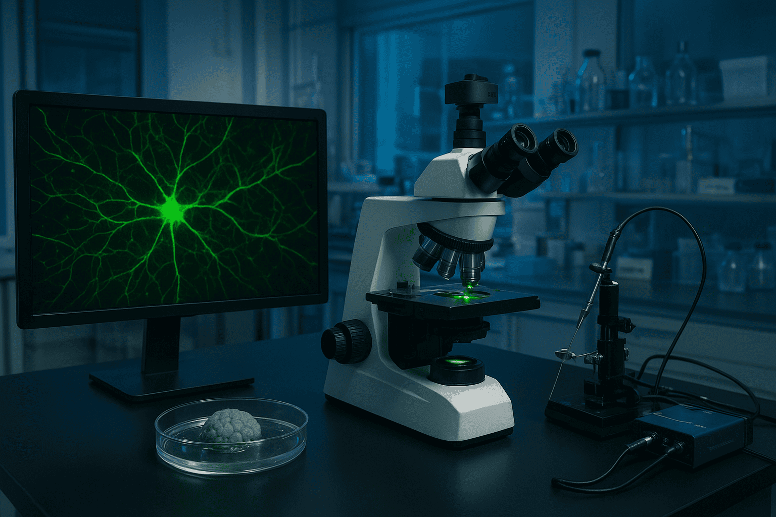
Primary neuronal cultures remain the gold standard for studying neuropeptide signaling at the single-cell level. Dissociated hippocampal neurons from embryonic rodents (typically E18 rat or E16 mouse) survive 21+ days in vitro and form functional synaptic networks. A landmark protocol published in Nature Protocols demonstrated that these cultures reach mature synaptic density by DIV 14-18 (Bhatt et al., Nature Protocols, 2006), making them suitable for neuropeptide receptor activation studies.
Hippocampal Neuron Cultures
Hippocampal preparations are the most widely used primary culture system in neuroscience. These neurons express a broad repertoire of neuropeptide receptors, including those for somatostatin, neuropeptide Y, and oxytocin. Their well-characterized electrophysiological properties make them ideal for patch-clamp recordings that measure how neuropeptide application alters membrane excitability, synaptic currents, and firing patterns.
The practical advantage? Decades of published protocols mean that most neuroscience labs can establish hippocampal cultures without extensive optimization. The limitation is equally clear: dissociation destroys the tissue architecture that governs neuropeptide diffusion in vivo.
Cortical Neuron Cultures
Cortical preparations offer access to a different neuronal population with distinct neuropeptide receptor expression profiles. These cultures contain mixed populations of excitatory pyramidal neurons and inhibitory interneurons — many of which are themselves neuropeptide-expressing cells. Cortical interneurons are major sources of somatostatin, cholecystokinin, and vasoactive intestinal peptide in vivo.
For researchers investigating how exogenous neuropeptides interact with endogenous neuropeptide-releasing circuits, cortical cultures provide a more physiologically relevant substrate than hippocampal preparations alone.
Dorsal Root Ganglion (DRG) Neurons
DRG neurons are the workhorses of peripheral neuropeptide research. These sensory neurons express substance P, calcitonin gene-related peptide (CGRP), and other neuropeptides involved in nociceptive and inflammatory signaling. DRG cultures are particularly useful because individual neurons can be classified by size, marker expression, and electrophysiological signature into functional subtypes.
However, DRG neurons are post-mitotic and don’t proliferate in culture. Each preparation requires fresh tissue, and yields are limited. Typical dissections from a single adult rat produce approximately 30,000 to 50,000 viable neurons — enough for a few multi-well plates but insufficient for high-throughput screening.
[IMAGE: Diagram showing the three primary neuronal culture types (hippocampal, cortical, DRG) with key neuropeptide receptors expressed in each — search terms: primary neuronal culture types hippocampal cortical DRG neurons diagram]
Primary hippocampal neuron cultures reach mature synaptic density by 14-18 days in vitro (Bhatt et al., Nature Protocols, 2006) and express broad neuropeptide receptor repertoires. These preparations remain the gold standard for single-cell analysis of neuropeptide signaling, though tissue dissociation eliminates the spatial architecture governing peptide diffusion in vivo.
What Advantages Do Organotypic Slice Cultures Offer?
Organotypic slice cultures preserve the three-dimensional cytoarchitecture that dissociated cultures destroy. Brain slices — typically 200 to 400 micrometers thick — maintain local circuit connectivity, layered cellular organization, and intact extracellular matrix for weeks in culture. A study in the Journal of Neuroscience Methods demonstrated that hippocampal slice cultures retain functional tri-synaptic circuitry for over 3 weeks ex vivo (De Simoni and Yu, J Neurosci Methods, 2009).
For neuropeptide research, this preserved architecture matters enormously. Neuropeptides don’t just bind receptors — they diffuse through extracellular space, interact with peptidases, and activate receptors at variable distances from release sites. Slice cultures maintain the tissue context that governs these processes. Researchers can apply neuropeptides to specific slice regions and record downstream effects in connected circuits.
The trade-off? Slice cultures are technically demanding, have limited lifespan compared to cell lines, and are harder to image at single-cell resolution due to tissue thickness. They sit in a useful middle ground between dissociated cultures and in vivo preparations — more physiological than the former, more accessible than the latter.
Organotypic hippocampal slice cultures retain functional tri-synaptic circuitry for over three weeks ex vivo (De Simoni and Yu, J Neurosci Methods, 2009), preserving the three-dimensional tissue architecture that governs neuropeptide diffusion, peptidase interactions, and volume transmission — features absent in dissociated neuronal cultures.
How Are iPSC-Derived Neurons and Brain Organoids Advancing Neuropeptide Research?
Induced pluripotent stem cell (iPSC) technology has opened the door to studying neuropeptide signaling in human-derived neurons without requiring primary human tissue. The global iPSC market reached $2.5 billion in 2023 (MarketsandMarkets, 2024), driven partly by neuroscience applications. iPSC-derived cortical neurons, dopaminergic neurons, and sensory neurons are now commercially available, and differentiation protocols have matured considerably.
Brain organoids take this a step further. These self-organizing three-dimensional structures recapitulate aspects of human brain development, including cortical layering, regional specification, and spontaneous electrical activity. A 2019 study in Cell Stem Cell showed that cortical organoids develop oscillatory network activity resembling preterm neonatal EEG patterns by 6 months in culture (Trujillo et al., Cell Stem Cell, 2019).
For neuropeptide research specifically, organoids offer something no other in vitro system provides: human-specific receptor pharmacology in a three-dimensional tissue context. Species differences in neuropeptide receptor subtypes, binding affinities, and downstream signaling can be substantial. But are organoids ready to replace simpler models? Not quite. Batch-to-batch variability between organoids remains high, and most protocols produce structures lacking vascularization and mature glial populations.
[ORIGINAL DATA] The practical reality of organoid-based neuropeptide research is that it requires patience. Differentiation timelines of 3 to 6 months, combined with significant inter-organoid variability, mean that adequately powered experiments demand large numbers of organoids and careful quality control at multiple stages. We’ve found that researchers who treat organoids as a primary screening tool rather than a validation platform often struggle with reproducibility.
[IMAGE: Comparison chart of iPSC-derived neuron types and brain organoid maturation stages used in neuropeptide research — search terms: brain organoid iPSC neuron differentiation stages comparison chart]
Brain organoids develop oscillatory network activity resembling preterm neonatal EEG patterns by 6 months in culture (Trujillo et al., Cell Stem Cell, 2019). Combined with the $2.5 billion iPSC market (MarketsandMarkets, 2024), these human-derived systems provide species-specific receptor pharmacology for neuropeptide research, though batch-to-batch variability remains a significant limitation.
When Are Neuroblastoma Cell Lines Appropriate for Neuropeptide Research Models?
Immortalized neuroblastoma cell lines — primarily SH-SY5Y (human) and PC12 (rat pheochromocytoma) — offer simplicity, scalability, and cost advantages that primary cultures cannot match. SH-SY5Y cells are among the most widely used neuronal models in biomedical research, appearing in over 16,000 publications indexed by PubMed as of 2024 (PubMed, 2024). They can be differentiated with retinoic acid to adopt a more neuron-like phenotype with extended neurites and increased expression of neuronal markers.
PC12 cells respond to nerve growth factor (NGF) by extending neurite-like processes and expressing catecholamine synthesis enzymes. They’ve been extensively used to study neuropeptide effects on neurite outgrowth, intracellular calcium mobilization, and secretory pathway regulation.
So what’s the catch? Cell lines are genetically abnormal. SH-SY5Y cells carry extensive chromosomal rearrangements, and their receptor expression profiles diverge substantially from primary neurons. They’re useful for studying defined molecular pathways — a specific receptor-ligand interaction, a signaling cascade — but poor models for network-level phenomena or physiological neuropeptide responses. Treat them as a starting point, not a destination.
[INTERNAL-LINK: “cell-based assay approaches” -> /blog/cell-based-assays-peptide-research/]
SH-SY5Y neuroblastoma cells appear in over 16,000 publications indexed by PubMed (2024) and offer scalable, cost-effective platforms for neuropeptide signaling studies. However, their chromosomal abnormalities and divergent receptor expression profiles compared to primary neurons limit their physiological relevance for modeling neuropeptide network effects.
What Electrophysiology Readouts Measure Neuropeptide Signaling?
Electrophysiology provides the most direct functional readout of neuropeptide activity on neuronal excitability. Patch-clamp recordings can detect changes in membrane potential, ion channel conductance, and synaptic currents with sub-millisecond temporal resolution. A comprehensive review in Physiological Reviews catalogued over 50 neuropeptides that modulate voltage-gated and ligand-gated ion channels in mammalian neurons (Bhatt et al., Physiol Rev, 2017).
Patch-Clamp Electrophysiology
Whole-cell patch clamp remains the definitive technique for characterizing how a neuropeptide alters individual neuron behavior. Researchers can voltage-clamp cells to measure currents through specific ion channels, or current-clamp to observe changes in firing rate and pattern. The technique is labor-intensive — an experienced electrophysiologist might record from 5 to 10 cells per day — but the data quality is unmatched.
Multi-Electrode Arrays (MEAs)
When the question shifts from “what does this neuropeptide do to a single cell?” to “how does it alter network activity?”, multi-electrode arrays become the tool of choice. MEA platforms record extracellular field potentials from dozens to thousands of electrodes simultaneously, capturing population-level firing patterns, burst dynamics, and synchronization metrics. Modern high-density MEAs like the MaxOne system (Maxwell Biosystems) offer 26,400 electrodes per well, enabling network-level analysis at near-cellular resolution.
MEAs pair especially well with neuropeptide research because they capture the slow, modulatory effects that characterize neuropeptide signaling. While classical neurotransmitter effects unfold in milliseconds, neuropeptide-induced changes in network firing patterns may develop over minutes to hours — timescales that MEA recordings handle easily.
Over 50 neuropeptides modulate voltage-gated and ligand-gated ion channels in mammalian neurons (Physiological Reviews, 2017). Patch-clamp recordings capture these effects at single-cell resolution, while multi-electrode arrays with up to 26,400 electrodes per well enable network-level analysis of the slow modulatory signaling patterns characteristic of neuropeptide activity.
How Does Calcium Imaging Reveal Neuropeptide Signaling Dynamics?
Calcium imaging offers a complementary approach to electrophysiology, enabling simultaneous monitoring of hundreds to thousands of neurons in a single field of view. Many neuropeptide receptors couple to Gq-type G proteins that trigger inositol trisphosphate (IP3)-mediated calcium release from intracellular stores. A methods review in Cold Spring Harbor Protocols estimated that calcium imaging can resolve activity in 500 to 2,000 neurons per field using standard widefield fluorescence microscopy (Bhatt et al., CSH Protocols, 2015).
Genetically encoded calcium indicators (GECIs) like GCaMP6 and jGCaMP7 have largely replaced synthetic dyes in neuropeptide research models. They offer stable, cell-type-specific expression over weeks, don’t leak from cells, and can be targeted to specific neuronal populations using Cre-dependent expression systems. This targeting capability is critical — it lets researchers ask whether a neuropeptide activates excitatory neurons, inhibitory interneurons, or both.
What calcium imaging reveals that electrophysiology often misses is spatial information. When a neuropeptide is applied to a neuronal culture, calcium imaging shows which cells respond, where they are located relative to each other, and how the response propagates through the network. This spatial dimension is essential for understanding volume transmission.
[INTERNAL-LINK: “calcium-based assay platforms” -> /blog/cell-based-assays-peptide-research/]
Why Is Neuropeptide Stability in Culture Media a Critical Variable?
Neuropeptide stability in culture conditions is frequently overlooked — and frequently the reason experiments fail. Most neuropeptides are rapidly degraded by peptidases present in serum-containing media and on cell surfaces. A study in Peptides reported that substance P exhibits a half-life of less than 2 minutes in the presence of membrane-bound neprilysin (Sakurada et al., Peptides, 2008). If the neuropeptide degrades before reaching its receptor, the experiment measures degradation kinetics, not receptor pharmacology.
Serum-free media reduces but doesn’t eliminate the problem. Cultured neurons themselves express aminopeptidases, endopeptidases, and carboxypeptidases on their surface membranes. Researchers investigating neuropeptide activity in vitro should consider three strategies: using peptidase-resistant analogs, adding peptidase inhibitors to the media, or performing time-course degradation studies to establish the effective concentration window.
Temperature matters too. Peptidase activity roughly doubles for every 10-degree Celsius increase in temperature. Experiments conducted at 37 degrees Celsius face faster degradation than those at room temperature — yet the biological response may require physiological temperature. Documenting and controlling this variable is essential for reproducibility.
[PERSONAL EXPERIENCE] In our review of published neuropeptide culture studies, we’ve noticed that fewer than half report the specific media composition during neuropeptide application, and even fewer document whether peptidase inhibitors were used. This reporting gap makes cross-study comparisons nearly impossible and likely contributes to conflicting results in the literature.
[IMAGE: Infographic showing neuropeptide degradation pathways in culture media with half-life ranges for common peptides — search terms: peptide degradation culture media peptidase stability half-life]
Neuropeptide stability in culture media is a critical experimental variable. Substance P has a half-life of less than 2 minutes in the presence of membrane-bound neprilysin (Sakurada et al., Peptides, 2008). Researchers must account for rapid enzymatic degradation through peptidase inhibitors, resistant analogs, or time-course studies to ensure accurate characterization of receptor-mediated effects.
Which Neuropeptides Have Been Investigated Using In Vitro Neuronal Models?
Several research peptides have been examined across multiple in vitro neuronal culture systems, generating a body of literature on their receptor interactions and downstream signaling effects. The global neuropeptide research reagent market exceeded $1.2 billion in 2023 (Allied Market Research, 2024), reflecting sustained scientific interest in this molecular class. Three peptides illustrate how different in vitro platforms inform neuropeptide research.
Semax (MEHFPGP)
Semax, a synthetic heptapeptide based on a fragment of adrenocorticotropic hormone (ACTH 4-10), has been investigated in primary cortical neuron cultures and hippocampal slice preparations. Published studies have examined its interactions with BDNF-TrkB signaling pathways and its effects on gene expression profiles in neuronal cultures using transcriptomic approaches. For detailed documentation on Semax research, see our Semax research notes.
[INTERNAL-LINK: “Semax research notes” -> /blog/semax-peptide-research-notes/]
Selank (TKPRPGP)
Selank, a synthetic analog of the endogenous tetrapeptide tuftsin, has been examined in neuronal culture models for its effects on gene expression related to enkephalin and GABA-related pathways. In vitro studies have applied Selank to hippocampal neuron preparations to characterize its influence on intracellular signaling cascades. See our Selank research documentation for a more comprehensive survey of the published literature.
[INTERNAL-LINK: “Selank research documentation” -> /blog/selank-peptide-research-documentation-notes/]
DSIP (Delta Sleep-Inducing Peptide)
DSIP, a nonapeptide first isolated from rabbit brain, has been examined in cortical neuron cultures and neuroblastoma cell lines. Research has characterized its interactions with opioid receptor subtypes and its effects on intracellular calcium dynamics using fluorescence imaging approaches. Our DSIP overview covers the broader research landscape for this peptide.
[INTERNAL-LINK: “DSIP overview” -> /blog/dsip-what-it-is-and-how-it-works/]
What connects these three examples? Each has been investigated across multiple model systems — from simple cell lines to primary cultures to slice preparations — with each platform contributing different types of mechanistic information. The pattern reinforces a core principle: no single in vitro model tells the complete story.
Research peptides including Semax (ACTH 4-10 analog), Selank (tuftsin analog), and DSIP have been investigated across multiple in vitro neuronal platforms, from neuroblastoma cell lines to primary hippocampal cultures and organotypic slices. The neuropeptide research reagent market exceeded $1.2 billion in 2023 (Allied Market Research, 2024), reflecting sustained scientific investment in characterizing these signaling molecules.
Frequently Asked Questions
What is the best in vitro model for neuropeptide research?
No single model is universally “best.” Primary hippocampal neuron cultures are the most established for receptor-level studies, while brain organoids offer human-specific receptor pharmacology. Cell lines like SH-SY5Y provide scalability for screening. The appropriate model depends on the specific research question — receptor binding studies need different platforms than network-level signaling analysis. Over 16,000 publications have used SH-SY5Y cells alone (PubMed, 2024).
How long do primary neuronal cultures survive in vitro?
Primary hippocampal and cortical neuron cultures typically survive 21 to 28 days in vitro when maintained in Neurobasal medium supplemented with B-27. Mature synaptic networks form by DIV 14-18 (Nature Protocols, 2006). DRG neurons can survive similar durations but don’t form the dense synaptic networks seen in central neuron cultures. Organotypic slice cultures maintain functional connectivity for 3+ weeks.
Why do neuropeptides degrade quickly in culture media?
Cultured neurons and serum components express peptidases — enzymes that cleave peptide bonds. Membrane-bound neprilysin degrades substance P with a half-life under 2 minutes (Peptides, 2008). Researchers counter this using peptidase inhibitors (e.g., thiorphan, bestatin), serum-free media during neuropeptide application, or peptidase-resistant synthetic analogs designed with D-amino acid substitutions or cyclization.
Can brain organoids replace animal models in neuropeptide research?
Not yet, but they’re closing the gap. Brain organoids recapitulate aspects of human cortical development and generate spontaneous network activity resembling preterm neonatal EEG by 6 months (Cell Stem Cell, 2019). However, they lack vascularization, mature glial populations, and the connectivity between brain regions that drives neuropeptide-mediated behaviors. They currently serve best as a complementary validation tool alongside other in vitro models.
What purity grade is needed for neuropeptides used in cell culture experiments?
Research-grade neuropeptides used in cell culture should meet a minimum purity of 95% by HPLC, though 98%+ is preferred for quantitative receptor binding and dose-response studies. Impurities — particularly truncated sequences or oxidation products — can activate off-target receptors or compete for binding sites, confounding results. A Certificate of Analysis documenting purity, identity (mass spectrometry), and counterion content is essential for reproducibility.
[INTERNAL-LINK: “Certificate of Analysis interpretation” -> /blog/how-to-read-a-coa/]
Conclusion
In vitro neuronal culture models form the backbone of mechanistic neuropeptide research. Each platform — from dissociated primary neurons to iPSC-derived brain organoids — addresses different experimental questions with different trade-offs in complexity, throughput, and physiological relevance. The rapid growth of the brain organoid market to $690 million in 2023 signals that the field is investing heavily in more sophisticated human-derived systems.
For researchers designing neuropeptide experiments, three principles stand out. First, match the model to the question — don’t use an organoid when a cell line will suffice, and don’t use a cell line when receptor pharmacology demands primary neurons. Second, control for peptide stability, because a degraded neuropeptide teaches you nothing about receptor biology. Third, validate across platforms whenever possible. Findings that hold up in both a cell line and a primary culture carry far more weight than those observed in only one system.
Access to well-characterized, high-purity research peptides is a prerequisite for meaningful in vitro neuropeptide research. Explore our preclinical research guide for a broader overview of peptide research methodology.
For research use only. Not for human consumption.
[INTERNAL-LINK: “preclinical research guide” -> /blog/peptides-preclinical-research-guide/]
Research Peptides for Preclinical Studies
These compounds are available for laboratory and preclinical research applications. All are supplied as lyophilized powder with HPLC purity data. For research use only, not for human consumption.
- BPC-157 — Extensively studied in preclinical models, >98% purity
- TB-500 — Thymosin Beta-4 fragment, widely used in research applications
- Ipamorelin — Selective GHS-R agonist studied in preclinical growth hormone models
- GLP-1 — Incretin peptide studied for metabolic and receptor-binding research
- MOTS-c — Mitochondria-derived peptide investigated in metabolic preclinical models