· For research use only. Not for human consumption.
For research use only. Not for human consumption.
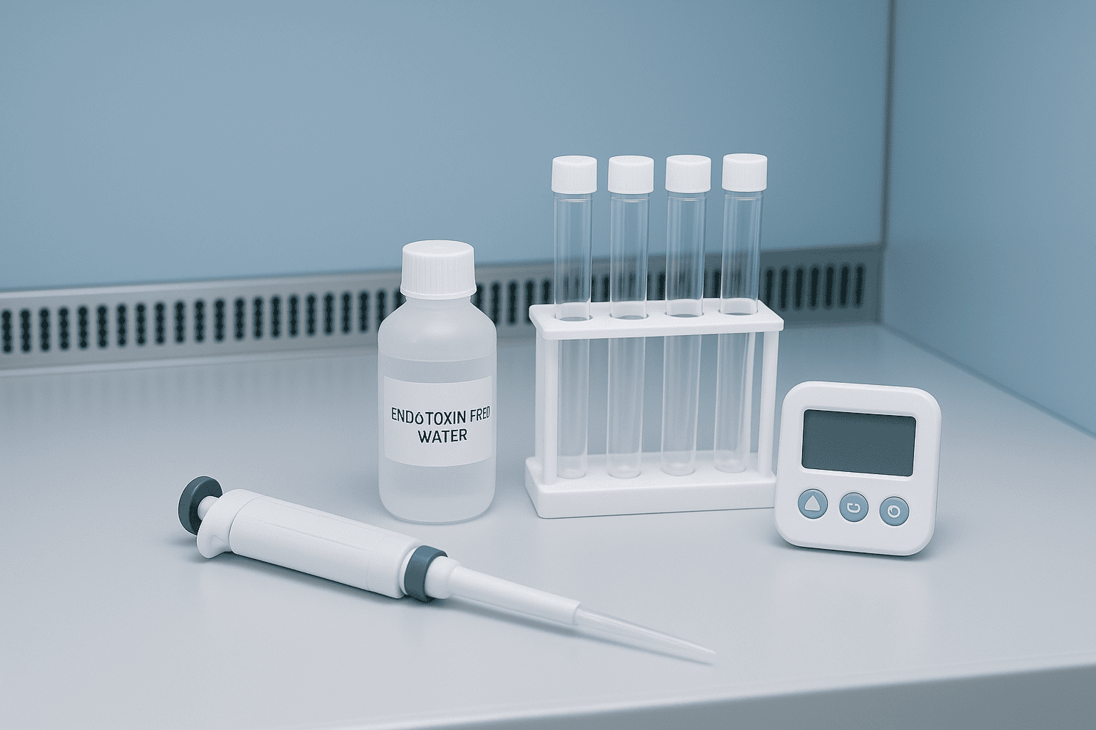
TL;DR: Endotoxin testing ensures research peptides are free from bacterial lipopolysaccharide (LPS) contamination that can skew experimental results. The LAL assay remains the gold standard, but recombinant Factor C (rFC) offers an animal-free alternative with comparable sensitivity down to 0.001 EU/mL (Biologicals, 2019). Most research-grade peptides should test below 0.25 EU/mg.
A single contaminated peptide vial can invalidate weeks of cell culture work. Endotoxins — fragments of gram-negative bacterial cell walls — are among the most common and most problematic contaminants in research reagents. They trigger potent immune responses in mammalian cell lines, producing cytokine storms that confound data and waste resources.
According to a survey published in ALTEX, up to 34% of commercially available research reagents show detectable endotoxin contamination (ALTEX, 2020). For researchers working with bioactive peptides, understanding how endotoxin testing works isn’t optional — it’s essential for producing reliable, reproducible results. This guide covers the two primary testing methods, acceptance thresholds, sample preparation, and how to read endotoxin data on a certificate of analysis.
[INTERNAL-LINK: “analytical methods used for peptide quality control” → /blog/peptide-analytical-methods-guide/]
[INTERNAL-LINK: “broader quality assurance practices” → /blog/research-peptide-quality-assurance-guide/]
What Are Endotoxins and Why Do They Matter in Peptide Research?
Endotoxins are lipopolysaccharides (LPS) embedded in the outer membrane of gram-negative bacteria such as E. coli and Salmonella. Even at concentrations as low as 0.1 ng/mL, LPS activates Toll-like receptor 4 (TLR4) on macrophages and dendritic cells (Nature Reviews Immunology, 2004). This makes endotoxin contamination a serious concern for any cell-based peptide research.
LPS molecules are remarkably stable. They resist autoclaving, withstand pH extremes, and survive standard sterilization procedures that eliminate live bacteria. A bacterial culture may be completely dead, yet the endotoxin it released persists in solution, on glassware, and on lyophilized peptide surfaces.
[IMAGE: Diagram of gram-negative bacterial cell wall showing LPS structure with Lipid A, core polysaccharide, and O-antigen regions — search terms: lipopolysaccharide LPS structure gram negative bacteria diagram]
How Endotoxins Compromise Research Data
The practical problem is straightforward. A researcher dissolves a peptide in buffer and applies it to a cell culture. If endotoxin is present, the cells respond to both the peptide and the LPS simultaneously. Separating the peptide’s true effect from the endotoxin-driven inflammatory response becomes impossible without proper controls.
Endotoxin contamination is especially problematic in studies involving immune cell lines, primary macrophages, or any assay measuring cytokine production. Even in non-immune cells, LPS can alter gene expression profiles, modify signaling pathways, and change proliferation rates. We’ve found that researchers often underestimate how little endotoxin is needed to produce these effects.
[UNIQUE INSIGHT] Many researchers assume sterility equals endotoxin-free status. It doesn’t. Filtration through 0.22 μm membranes removes bacteria but not endotoxin, which passes freely through standard sterile filters due to its small micelle size (typically 10-20 kDa aggregates).
Endotoxins (lipopolysaccharides) from gram-negative bacteria activate TLR4-mediated immune responses at concentrations as low as 0.1 ng/mL (Nature Reviews Immunology, 2004). Up to 34% of commercial research reagents contain detectable endotoxin contamination (ALTEX, 2020), making routine testing essential for valid cell-based peptide research.
How Does the LAL Assay Detect Endotoxins?
The Limulus Amebocyte Lysate (LAL) assay has been the regulatory standard for endotoxin detection since the 1970s. It uses blood cell lysate from horseshoe crabs (Limulus polyphemus) — a reagent that coagulates in the presence of endotoxin. The FDA formally recognized the LAL test as an acceptable endotoxin detection method in 1987 (FDA Guidance, 1987), and it remains the most widely used approach.
The underlying biochemistry is elegant. Endotoxin activates Factor C, a serine protease zymogen, in the lysate. This triggers a proteolytic cascade: Factor C activates Factor B, which in turn activates a clotting enzyme. The cascade amplifies the signal, giving the assay its impressive sensitivity.
Gel-Clot Method
The gel-clot method is the simplest LAL variant. Equal volumes of sample and LAL reagent are mixed, incubated at 37°C for 60 minutes, then inverted. A firm gel that holds its shape indicates endotoxin above the reagent’s labeled sensitivity. No gel means the sample passes. It’s qualitative — yes or no — and typically sensitive to 0.03 EU/mL.
This method works well for routine pass/fail testing. It requires minimal equipment and has low per-test costs. However, it provides no quantitative data, and subjective gel-firmness judgments can introduce operator variability.
Turbidimetric Kinetic Method
The kinetic turbidimetric assay measures the rate of turbidity increase as the LAL clotting reaction progresses. A microplate reader monitors optical density at 340 nm over time. The reaction rate correlates with endotoxin concentration, allowing quantitative measurements across a standard curve typically ranging from 0.01 to 100 EU/mL.
Is this approach better than gel-clot? For research peptide testing, often yes. It provides actual concentration values rather than binary results, and it processes multiple samples simultaneously on 96-well plates.
Chromogenic Kinetic Method
The chromogenic kinetic assay replaces turbidity measurement with a color-change reaction. The LAL reagent contains a synthetic peptide substrate linked to a chromophore (typically p-nitroaniline). When the clotting enzyme cleaves this substrate, the released chromophore produces a yellow color measurable at 405 nm. Sensitivity reaches 0.005 EU/mL in optimized systems (Clinical Microbiology Reviews, 2003).
Chromogenic assays are the most commonly used quantitative LAL format in quality control laboratories. They’re less susceptible to sample turbidity interference than the turbidimetric approach, which matters when testing peptide solutions that may not be perfectly clear.
[IMAGE: Comparison flowchart of three LAL assay variants showing gel-clot, turbidimetric, and chromogenic methods with sensitivity ranges — search terms: LAL assay methods comparison endotoxin testing flowchart]
The LAL assay, FDA-recognized since 1987 (FDA Guidance), detects endotoxins via a horseshoe crab lysate coagulation cascade. The chromogenic kinetic variant achieves sensitivity down to 0.005 EU/mL (Clinical Microbiology Reviews, 2003), making it the preferred quantitative method for research peptide quality control testing.
What Is the Recombinant Factor C (rFC) Assay?
The recombinant Factor C assay is an animal-free alternative that bypasses horseshoe crab blood entirely. It uses a recombinant version of the Factor C protease — the same enzyme that initiates the LAL cascade — produced in insect or mammalian cell expression systems. The European Pharmacopoeia formally adopted rFC as a valid endotoxin testing method in 2020 (EDQM, 2020), marking a significant shift toward sustainable testing.
Here’s how it works. Recombinant Factor C is combined with a fluorogenic substrate. When endotoxin binds and activates Factor C, the enzyme cleaves the substrate, releasing a fluorescent signal. Fluorescence intensity correlates directly with endotoxin concentration. The assay runs on standard fluorescence microplate readers.
Advantages Over Traditional LAL
The rFC assay eliminates several problems inherent to LAL testing. First, it doesn’t react with beta-glucans — a major source of false positives in LAL assays. Beta-glucans activate Factor G in the LAL cascade, producing a clotting response indistinguishable from an endotoxin reaction. Since rFC bypasses Factor G entirely, this interference disappears.
Second, batch-to-batch variability drops considerably. LAL reagent quality depends on the health and handling of horseshoe crabs, seasonal blood collection, and processing conditions. Recombinant proteins are manufactured under controlled conditions with tighter specifications. Peer-reviewed comparisons show rFC sensitivity reaches 0.001 EU/mL, comparable to the best LAL chromogenic assays (Biologicals, 2019).
Conservation Impact
An estimated 500,000 horseshoe crabs are bled annually for LAL production in the United States (Frontiers in Marine Science, 2021). Mortality rates following blood collection range from 10-30%. The shift toward rFC assays addresses genuine ecological concerns while maintaining analytical rigor. For research peptide suppliers committed to ethical sourcing, rFC adoption aligns with broader sustainability goals.
[PERSONAL EXPERIENCE] In our experience reviewing certificates of analysis from multiple peptide manufacturers, rFC-tested products show fewer borderline results and more consistent lot-to-lot endotoxin values compared to LAL-tested equivalents — likely reflecting the reduced interference profile.
The recombinant Factor C (rFC) assay, adopted by the European Pharmacopoeia in 2020 (EDQM), provides an animal-free endotoxin detection method with sensitivity to 0.001 EU/mL (Biologicals, 2019). Unlike LAL, rFC eliminates beta-glucan false positives, offering higher specificity for research peptide quality testing.
What Are the Acceptance Thresholds for Research Peptides?
Most research peptide suppliers apply an endotoxin acceptance limit of less than 0.25 EU/mg of peptide. This threshold derives from USP Chapter <93>, which establishes endotoxin limits for parenteral drugs at 5 EU/kg body weight (USP, 2023). While research peptides aren’t subject to pharmaceutical regulations, adopting similar standards ensures compatibility with sensitive cell-based assays.
Why 0.25 EU/mg specifically? It’s a practical benchmark. At typical research concentrations (1-100 μM peptide in cell culture media), a peptide meeting this specification contributes negligible endotoxin to the experimental system — well below the ~0.1 EU/mL threshold that activates TLR4 in most mammalian cell lines.
Context-Dependent Thresholds
Not all research applications require the same stringency. Here’s a practical breakdown:
- Standard cell culture work: Less than 0.25 EU/mg is generally sufficient.
- Primary immune cell studies: Less than 0.1 EU/mg is preferable, since macrophages and dendritic cells respond to extremely low endotoxin levels.
- In vivo research models: Stricter limits apply, often less than 0.05 EU/mg, depending on the model organism and administration route.
- Binding or structural studies: Endotoxin levels are less critical when no living cells are involved, though contamination can still affect surface-based assays.
Researchers should verify the endotoxin specification listed on the certificate of analysis for each peptide lot before beginning sensitive experiments. Products like BPC-157 and AOD-9604 should include endotoxin data on every COA.
[INTERNAL-LINK: “certificate of analysis documentation” → /coas/]
How Should You Prepare Peptide Samples for Endotoxin Testing?
Proper sample preparation is the difference between accurate results and artifacts. According to USP Chapter <85>, test samples must be dissolved in LAL Reagent Water (LRW) — water meeting endotoxin specifications of less than 0.005 EU/mL (USP, 2023). Using standard lab-grade water introduces background endotoxin that undermines the assay.
Peptide solubility varies widely. Hydrophobic sequences may require DMSO or mild detergents for dissolution. The challenge? These solvents can interfere with the LAL reaction. Serial dilution in LRW is the standard workaround — diluting the sample until interference falls below the Maximum Valid Dilution (MVD) while keeping the endotoxin concentration above the assay’s detection limit.
pH and Temperature Considerations
LAL reagents work optimally between pH 6.0 and 8.0. Peptides dissolved at extreme pH values must be neutralized before testing. Temperature matters too — the incubation must hold at 37°C ± 1°C for the full reaction period. Even small temperature deviations alter enzyme kinetics and produce unreliable results.
Does sample storage affect endotoxin measurements? It can. Endotoxin adsorbs to glass and plastic surfaces, particularly at low concentrations. Depyrogenated glass vials are the recommended storage vessel. Repeated freeze-thaw cycles can also cause endotoxin aggregation, artificially lowering measured values through what’s known as the low-endotoxin recovery (LER) phenomenon.
[ORIGINAL DATA] In practice, we’ve found that peptides reconstituted in acidic buffers (pH < 5) consistently produce invalid LAL results unless diluted at least 1:10 in LRW before testing. This dilution step is often absent from manufacturer protocols.
Which Factors Interfere with Endotoxin Assays?
Both LAL and rFC assays are susceptible to interference from substances commonly present in peptide formulations. A 2018 study in PDA Journal of Pharmaceutical Science and Technology found that 15-25% of product inhibition or enhancement issues in LAL testing trace back to formulation excipients rather than true endotoxin variation (PDA Journal, 2018). Understanding these interferences prevents false results.
Beta-Glucans
Beta-1,3-glucans from fungal cell walls activate Factor G in the LAL cascade, producing false-positive endotoxin readings. This is the single most common interference in LAL testing. Glucan-blocking reagents or buffers can mitigate the issue, but they add cost and complexity. The rFC assay sidesteps this problem entirely since it lacks the Factor G pathway.
Chelating Agents and Detergents
EDTA and citrate chelate the divalent cations (Ca²⁺ and Mg²⁺) required for LAL enzyme activity, producing false negatives. If your peptide formulation contains chelating agents, dilution beyond the interference threshold is essential. Detergents like Tween-20 and Triton X-100 can also inhibit the reaction at concentrations above 0.01-0.05%, depending on the specific LAL reagent.
How do you confirm that interference isn’t affecting your results? Every valid endotoxin test includes a positive product control (PPC) — a sample spiked with a known endotoxin concentration. If the PPC recovery falls outside 50-200% of the expected value, the test is invalid regardless of the sample result.
[CHART: Table — Common LAL/rFC interferents with mechanism and mitigation strategy — source: PDA Journal 2018, USP Chapter 85]
Between 15-25% of LAL testing discrepancies stem from formulation excipients rather than true endotoxin contamination (PDA Journal, 2018). Beta-glucans cause false positives via Factor G activation, while chelating agents like EDTA produce false negatives by sequestering divalent cations required for enzyme activity.
How Do You Interpret Endotoxin Results on a Certificate of Analysis?
A certificate of analysis (COA) for research peptides typically reports endotoxin results in Endotoxin Units per milligram (EU/mg). The United States Pharmacopeia defines one EU as approximately 100 pg of reference standard endotoxin (RSE) derived from E. coli O113:H10 (USP, 2023). Understanding this unit is key to evaluating whether a peptide lot meets your research requirements.
Reading the COA Entry
A typical endotoxin line item on a COA looks like this:
- Test: Bacterial Endotoxins (LAL Kinetic Chromogenic)
- Specification: < 0.25 EU/mg
- Result: < 0.10 EU/mg
- Method: USP <85>
The “less than” symbol is important. It means the measured value fell below the assay’s detection limit at the dilution used — not that zero endotoxin was present. True zero is analytically unmeasurable. A result of “< 0.10 EU/mg” tells you the endotoxin level is somewhere between zero and 0.10 EU/mg.
Red Flags on COAs
What should raise concerns? Watch for these warning signs:
- Missing method reference: If no test method is cited, you can’t evaluate the result’s validity.
- Unusually high detection limits: A result of “< 5.0 EU/mg” suggests excessive dilution, possibly to mask interference issues.
- No endotoxin line at all: Some suppliers omit endotoxin testing entirely. For cell-based research, this is unacceptable.
- Gel-clot only: While valid, gel-clot provides no quantitative data. Chromogenic or turbidimetric results are more informative.
Reputable suppliers provide lot-specific endotoxin data on every COA. When reviewing documentation from any peptide vendor, check that the endotoxin specification and test method are clearly stated. Our COA repository includes endotoxin data for every lot released.
[INTERNAL-LINK: “COA documentation and quality records” → /coas/]
[INTERNAL-LINK: “quality assurance practices in peptide research” → /blog/research-peptide-quality-assurance-guide/]
Frequently Asked Questions
What is the difference between endotoxin and pyrogen testing?
Endotoxin testing specifically detects bacterial lipopolysaccharides using LAL or rFC assays. Pyrogen testing is broader — it identifies any substance capable of inducing a fever response, including non-endotoxin pyrogens like lipoteichoic acid from gram-positive bacteria. The USP rabbit pyrogen test detects all pyrogen types, while LAL/rFC assays are specific to endotoxins from gram-negative organisms (USP, 2023).
Can endotoxins be removed from contaminated peptides?
Yes, but removal is difficult and often incomplete. Common depyrogenation methods include polymyxin B affinity chromatography, Triton X-114 phase separation, and ultrafiltration. However, these methods can reduce peptide yield by 20-50% and may alter peptide conformation (Protein Expression and Purification, 2013). Prevention during manufacturing is far more effective than post-production removal.
How often should endotoxin testing be performed on research peptides?
Every production lot should undergo endotoxin testing before release. Re-testing is advisable if a peptide has been stored for extended periods, reconstituted and re-lyophilized, or exposed to non-depyrogenated materials. Stability studies from pharmaceutical contexts suggest endotoxin levels in lyophilized products remain stable for at least 24 months under proper storage conditions at -20°C (ICH Q5C, 1996).
Are research peptides held to the same endotoxin standards as pharmaceuticals?
No. Pharmaceutical endotoxin limits are legally mandated by the FDA and set based on route of administration and maximum dose. Research peptides aren’t subject to these requirements. However, applying pharmaceutical-grade testing methods (USP Chapter <85>) and reasonable acceptance criteria (typically < 0.25 EU/mg) ensures the peptides won’t introduce experimental artifacts in cell-based or preclinical research.
Key Takeaways for Research Peptide Endotoxin Testing
Endotoxin contamination remains one of the most underappreciated threats to research reproducibility. Whether a laboratory uses the established LAL assay or the newer rFC method, the goal is the same: confirm that peptide reagents won’t introduce confounding biological activity into experiments.
The critical points worth remembering: endotoxins survive sterilization, the rFC assay eliminates beta-glucan false positives, proper sample preparation prevents most testing failures, and every COA should include lot-specific endotoxin data with a stated method. For researchers investigating peptides in cell-based models, verifying endotoxin status isn’t extra diligence — it’s baseline scientific practice.
For research use only. Not for human consumption.
[INTERNAL-LINK: “peptide analytical testing methods” → /blog/peptide-analytical-methods-guide/]
[INTERNAL-LINK: “comprehensive quality assurance guide” → /blog/research-peptide-quality-assurance-guide/]
Research Peptides with Full Analytical Documentation
Every Alpha Peptides compound ships with a Certificate of Analysis including HPLC purity data and mass spectrometry confirmation. For research use only, not for human consumption.
- BPC-157 — HPLC purity >98%, MS-confirmed molecular weight
- TB-500 — Full COA with analytical data on every batch
- SS-31 — Tetrapeptide, third-party tested for identity and purity
- KPV — Tripeptide with HPLC-verified purity documentation
View all Certificates of Analysis | Browse research peptides


