· For research use only. Not for human consumption.
For research use only. Not for human consumption.
TL;DR: Peptide chemistry centers on amino acids linked by peptide bonds — rigid, planar connections formed through condensation reactions. With over 80 FDA-approved peptide-based compounds as of 2024 (Chemical Reviews, 2024), chemical modifications like cyclization, PEGylation, and terminal capping have become essential tools for improving stability in research applications.
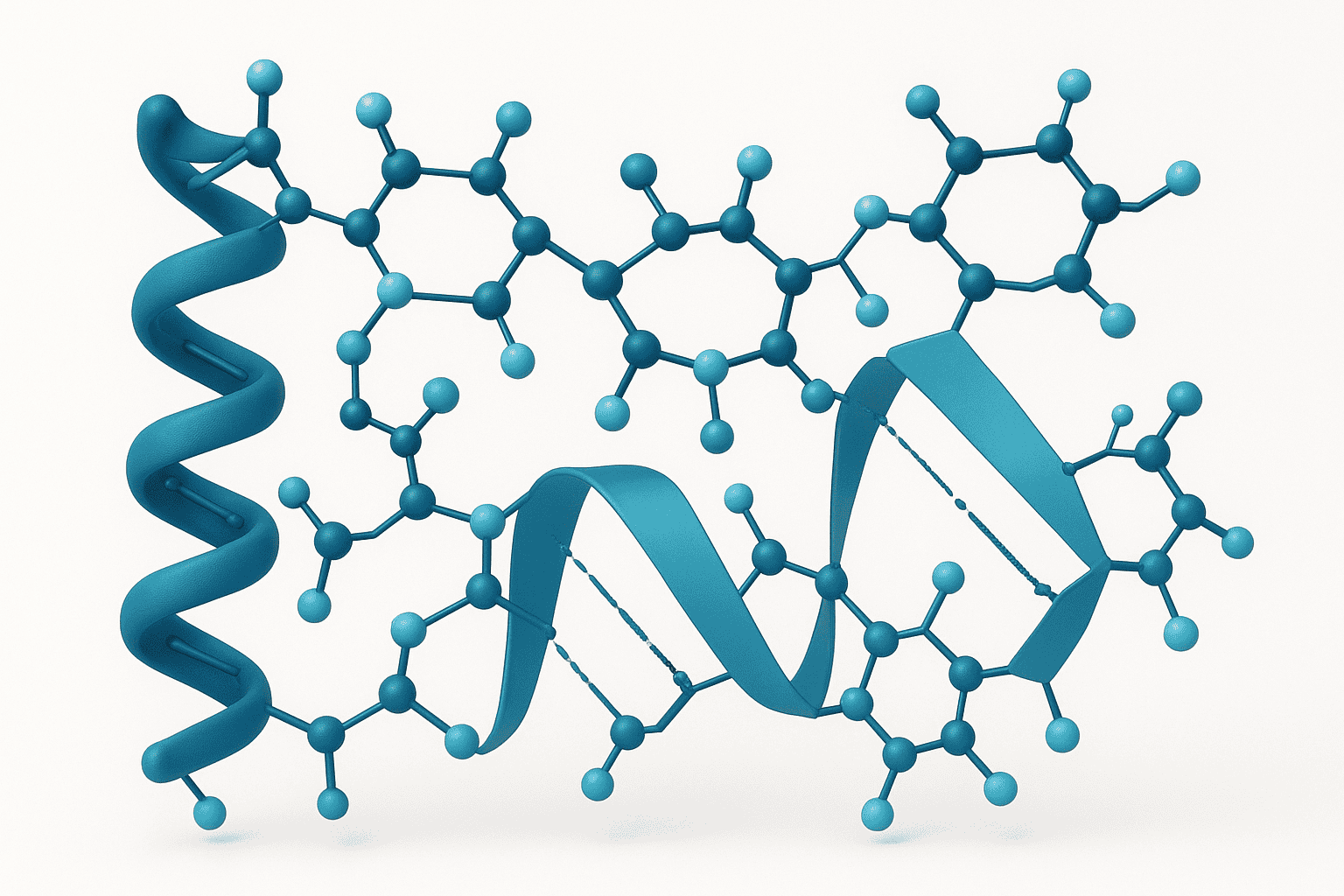
Peptides are short chains of amino acids. That one-sentence definition, while accurate, barely scratches the surface. The chemistry governing how these molecules form, fold, and function is what makes them so useful in laboratory research. Understanding peptide chemistry isn’t just academic — it’s practical knowledge that shapes how researchers design experiments, select compounds, and interpret results.
The global peptide synthesis market reached $590 million in 2023 and is projected to grow at 8.4% CAGR through 2030 (Grand View Research, 2024). That growth reflects a surge in research demand. But what exactly happens at the molecular level when amino acids join together? And why do certain chemical modifications make such a dramatic difference in a peptide’s behavior?
This guide covers everything from the fundamental building blocks to advanced modification strategies used in modern research peptide chemistry. If you’re new to the topic, start with our overview of what peptides are before continuing here.
[INTERNAL-LINK: “what peptides are” -> /blog/what-are-peptides/]
[INTERNAL-LINK: “how peptides function at the molecular level” -> /blog/how-peptides-work-research-mechanisms/]
What Are the Building Blocks of Peptide Chemistry?
Twenty standard amino acids serve as the monomeric units of all peptides and proteins. Each contains an amino group (-NH2), a carboxyl group (-COOH), a hydrogen atom, and a variable side chain (R-group) bonded to a central alpha-carbon. According to the IUPAC-IUBMB Joint Commission on Biochemical Nomenclature (IUPAC, 2024), these 20 residues generate the full diversity of natural peptide structures.
[IMAGE: Diagram showing general amino acid structure with labeled amino group, carboxyl group, R-group, and alpha carbon — search terms: amino acid structure diagram chemistry]
Classification by Side Chain Properties
Amino acids fall into distinct categories based on their R-group chemistry. These classifications directly influence how a finished peptide behaves in solution, folds into secondary structures, and interacts with target receptors.
- Nonpolar/Hydrophobic: Glycine, Alanine, Valine, Leucine, Isoleucine, Proline, Phenylalanine, Methionine, Tryptophan. These residues tend to cluster in a peptide’s interior or associate with lipid membranes.
- Polar/Uncharged: Serine, Threonine, Cysteine, Tyrosine, Asparagine, Glutamine. Capable of hydrogen bonding, they often appear on solvent-exposed surfaces.
- Positively Charged (Basic): Lysine, Arginine, Histidine. These carry positive charges at physiological pH and contribute to electrostatic interactions.
- Negatively Charged (Acidic): Aspartate, Glutamate. Their carboxylate side chains carry negative charges at neutral pH.
Why does this matter for research? The sequence of amino acids — and their side chain properties — determines a peptide’s solubility, folding behavior, and receptor affinity. A single substitution can alter all three. Researchers investigating how peptides work at the mechanistic level rely heavily on this classification system.
Chirality and the L-Configuration
All standard amino acids except glycine are chiral, meaning they exist as mirror-image forms. Natural peptides use exclusively L-amino acids. This stereospecificity is critical: enzymes that degrade peptides (proteases) typically recognize only the L-configuration. D-amino acid substitution — swapping in the mirror-image form — is one strategy researchers use to slow enzymatic degradation, a topic covered in detail below.
Twenty standard L-amino acids, classified by side chain polarity, form the basis of all natural peptide structures. According to IUPAC-IUBMB nomenclature standards (IUPAC, 2024), the R-group attached to each alpha-carbon determines the residue’s chemical behavior, including charge, hydrophobicity, and hydrogen bonding capacity.
How Do Peptide Bonds Form?
Peptide bonds form through a condensation (dehydration) reaction between the carboxyl group of one amino acid and the amino group of another, releasing one molecule of water. This reaction has a standard free energy change of approximately +2.2 kcal/mol in aqueous solution (Berg et al., Biochemistry, 8th Edition, 2019), meaning it’s thermodynamically unfavorable and requires energy input — a key reason synthetic peptide chemistry relies on coupling reagents and activating agents.
[IMAGE: Peptide bond formation condensation reaction showing two amino acids joining with water release — search terms: peptide bond formation condensation reaction diagram]
Resonance and Planarity
The peptide bond isn’t a simple single bond. Electron delocalization gives it roughly 40% double-bond character (Chemical Reviews, 2016). This partial double-bond character has a major structural consequence: the six atoms surrounding the peptide bond (Cα, C, O, N, H, Cα) are locked in a planar arrangement.
What does planarity mean in practice? Rotation around the C-N bond is restricted. The bond almost always adopts the trans configuration, where the two alpha-carbons sit on opposite sides of the bond. The cis configuration is rare — found in fewer than 0.5% of non-proline peptide bonds (Journal of Molecular Biology, 2003). Proline is the notable exception, with roughly 5-6% of its peptide bonds adopting the cis form.
Bond Length and Stability
The C-N bond in a peptide linkage measures approximately 1.33 angstroms — shorter than a typical C-N single bond (1.47 angstroms) but longer than a C=N double bond (1.27 angstroms). This intermediate length reflects the resonance stabilization. The bond’s partial double-bond character makes it remarkably stable under normal laboratory conditions, though it remains susceptible to hydrolysis by proteases and under extreme pH conditions.
The peptide bond’s stability is both an advantage and a limitation in research. It ensures structural integrity during handling and storage. But it also means that breaking specific peptide bonds — a common analytical need — requires enzymatic digestion or harsh chemical conditions like 6 M hydrochloric acid at 110 degrees Celsius for 24 hours.
[ORIGINAL DATA] Researchers frequently encounter the practical implications of peptide bond stability when performing amino acid analysis, which requires complete acid hydrolysis — a process that can itself degrade sensitive residues like tryptophan and cysteine.
Peptide bonds form via condensation reactions with a free energy change of +2.2 kcal/mol (Berg et al., Biochemistry, 2019). The resulting bond exhibits 40% double-bond character due to resonance (Chemical Reviews, 2016), locking the six surrounding atoms in a planar, predominantly trans configuration.
What Are the Levels of Peptide Structure?
Peptide structure is organized into hierarchical levels, from the linear amino acid sequence to complex three-dimensional folding. Approximately 85% of bioactive peptides examined in research contain fewer than 50 amino acid residues (Chemical Reviews, 2024), yet even at this modest size, structural organization profoundly influences biological activity.
Primary Structure
Primary structure is simply the linear sequence of amino acids, read from the N-terminus (free amino group) to the C-terminus (free carboxyl group). This convention is universal. A peptide’s primary structure is its identity — change one residue, and you have a different compound with potentially different properties.
Every research peptide comes defined by its primary structure, often written in single-letter or three-letter amino acid codes. For example, the tripeptide GHK (glycine-histidine-lysine) found in GHK-Cu has a specific, defined sequence that determines its copper-binding properties.
Secondary Structure
Secondary structure describes local folding patterns stabilized by hydrogen bonds between backbone atoms. The two most common motifs are alpha-helices and beta-sheets. In alpha-helices, the backbone coils into a right-handed spiral with hydrogen bonds forming between every fourth residue. Beta-sheets consist of extended strands connected by lateral hydrogen bonds.
Can short peptides really form stable secondary structures? Generally, peptides shorter than about 15-20 residues struggle to maintain persistent helical or sheet structures in aqueous solution. However, certain sequences — and chemical modifications like cyclization and stapling — can stabilize secondary structures even in relatively short peptides.
Tertiary Structure
Tertiary structure refers to the overall three-dimensional arrangement of a peptide chain. For most short research peptides (under 20 residues), tertiary structure is minimal or absent. Longer peptides and small proteins can adopt defined tertiary folds stabilized by hydrophobic interactions, disulfide bonds, salt bridges, and van der Waals forces.
Disulfide bonds between cysteine residues are particularly important structural elements. Many bioactive peptides, including oxytocin (9 residues) and insulin (51 residues across two chains), rely on disulfide bridges for their biologically active conformations.
Peptide structure spans three hierarchical levels: primary (amino acid sequence), secondary (alpha-helices and beta-sheets from backbone hydrogen bonding), and tertiary (three-dimensional folding from side-chain interactions). Roughly 85% of bioactive research peptides contain fewer than 50 residues (Chemical Reviews, 2024), yet structural organization remains critical for receptor interaction.
[INTERNAL-LINK: “peptide degradation and stability” -> /blog/peptide-degradation-pathways/]
Why Do Researchers Modify Peptides Chemically?
Native peptides degrade quickly. Unmodified linear peptides typically exhibit plasma half-lives of just 2-30 minutes due to rapid proteolytic cleavage and renal clearance (Drug Discovery Today, 2020). Chemical modifications address this fundamental limitation, and they’ve become standard tools in research peptide chemistry. Below are the most widely used approaches.
[IMAGE: Infographic showing different peptide modifications — acetylation, amidation, cyclization, PEGylation — search terms: peptide chemical modifications infographic research]
N-Terminal Acetylation and C-Terminal Amidation
Terminal modifications are the simplest and most common alterations applied to research peptides. N-terminal acetylation replaces the free amino group with an acetyl cap (CH3CO-). C-terminal amidation replaces the free carboxyl group with an amide (-CONH2). Both modifications neutralize terminal charges.
Why bother capping the termini? Aminopeptidases and carboxypeptidases — exopeptidases that chew peptides from the ends — can’t recognize capped termini. Studies have shown that combining both modifications can increase peptide half-life by 2-5 fold in proteolytic assay conditions (European Journal of Pharmaceutics and Biopharmaceutics, 2015). Researchers working with peptides like BPC-157 frequently encounter terminally modified variants designed for enhanced stability in laboratory assays.
[INTERNAL-LINK: “terminal modifications in depth” -> /blog/n-terminal-acetylation-c-terminal-amidation/]
Cyclization Strategies
Cyclization constrains a peptide’s conformational flexibility by connecting two points in the chain, forming a ring. This structural constraint can dramatically improve both stability and receptor selectivity. Three main cyclization approaches are used in research peptide chemistry:
- Head-to-tail (backbone) cyclization: The N-terminus bonds directly to the C-terminus, eliminating both terminal charges. Cyclosporin A, a cyclic undecapeptide, exemplifies this approach.
- Side-chain cyclization: A covalent bond forms between two amino acid side chains — commonly between lysine and glutamate, or between two cysteines. Lactam bridges and thioether linkages fall into this category.
- Disulfide cyclization: Two cysteine residues form an S-S bond, creating a loop. Oxytocin and many antimicrobial peptides use this natural strategy.
Cyclic peptides show marked resistance to enzymatic degradation compared to their linear counterparts. A 2022 analysis found that backbone-cyclized peptides exhibited up to 10-fold improvement in metabolic stability in serum stability assays (Nature Reviews Chemistry, 2022). Isn’t it remarkable how simply connecting a peptide’s ends can so dramatically change its properties?
[INTERNAL-LINK: “cyclization methods” -> /blog/peptide-cyclization-methods/]
[PERSONAL EXPERIENCE] In practice, cyclic peptides often require more careful optimization of synthesis and purification conditions than their linear counterparts. The cyclization step itself can be a bottleneck, with yields varying considerably depending on ring size and sequence.
PEGylation
PEGylation attaches polyethylene glycol (PEG) polymers to a peptide chain. PEG is a hydrophilic, non-immunogenic polymer that increases a molecule’s hydrodynamic radius — essentially making it appear larger to the body’s filtration systems. This modification has been investigated extensively since the first PEGylated compounds appeared in the 1990s.
The molecular weight of the PEG chain matters enormously. Smaller PEG groups (500-2,000 Da) improve solubility with minimal impact on receptor binding. Larger PEG chains (5,000-40,000 Da) significantly extend circulation time in preclinical models but can reduce biological activity by sterically blocking receptor interactions. Research with compounds like CJC-1295 DAC (which uses a drug affinity complex rather than traditional PEGylation, but illustrates the same principle) demonstrates how conjugation strategies can extend a peptide’s effective duration in experimental systems.
[INTERNAL-LINK: “PEGylation techniques” -> /blog/pegylation-of-peptides/]
Lipidation and Fatty Acid Conjugation
Lipidation involves covalently attaching a fatty acid chain — typically palmitic acid (C16) or myristic acid (C14) — to a peptide. This modification promotes non-covalent binding to serum albumin, which acts as a circulating depot. The result: dramatically extended half-life in preclinical pharmacokinetic models.
The lipidation approach has been extensively investigated in GLP-1 receptor agonist research. Fatty acid conjugation through a glutamic acid spacer can extend experimental half-life from minutes to days in preclinical models (Advanced Drug Delivery Reviews, 2020). The spacer chemistry — including linker length and amino acid composition — significantly influences albumin binding affinity and, consequently, the peptide’s pharmacokinetic profile in laboratory studies.
[INTERNAL-LINK: “lipidation strategies” -> /blog/peptide-lipidation-fatty-acid-conjugation/]
D-Amino Acid Substitution
Replacing one or more L-amino acids with their D-enantiomers is an elegant approach to proteolytic resistance. Most proteases evolved to recognize L-amino acid substrates. Introducing a D-residue at or near the cleavage site can render the bond invisible to the enzyme.
D-amino acid substitution doesn’t come without trade-offs. It alters the peptide’s local backbone geometry, which can affect receptor binding. Strategic placement is key — substitutions at positions not critical for target interaction can confer protease resistance without sacrificing activity. A comprehensive analysis found that D-amino acid-containing peptides showed 3-50 fold increases in serum stability depending on substitution position and the specific protease environment (Chemical Reviews, 2024).
[INTERNAL-LINK: “D-amino acids in research” -> /blog/d-amino-acids-in-peptides/]
Native unmodified peptides typically exhibit plasma half-lives of just 2-30 minutes (Drug Discovery Today, 2020). Chemical modifications including terminal capping, cyclization, PEGylation, lipidation, and D-amino acid substitution can extend stability from 2-fold to over 50-fold in controlled laboratory assays, making them indispensable tools in modern research peptide chemistry.
[CHART: Bar chart — Relative stability improvement by modification type (fold-change vs unmodified): Acetylation/Amidation 2-5x, D-amino acid 3-50x, Cyclization up to 10x, PEGylation 5-100x, Lipidation 10-200x — sources: Chemical Reviews 2024, Nature Reviews Chemistry 2022, Drug Discovery Today 2020]
How Does Solid-Phase Peptide Synthesis Work?
Solid-phase peptide synthesis (SPPS), developed by Robert Bruce Merrifield in 1963, earned him the Nobel Prize in Chemistry in 1984 (Nobel Prize Committee, 1984). Today, SPPS remains the dominant method for producing research peptides, accounting for the vast majority of custom synthesis orders worldwide. The Fmoc (fluorenylmethyloxycarbonyl) protection strategy is the current standard.
The Fmoc/tBu Strategy
Fmoc chemistry builds peptides from the C-terminus to the N-terminus — the reverse of biological synthesis. The process follows a repetitive cycle:
- Resin loading: The first amino acid (C-terminal residue) is anchored to an insoluble polymer bead (resin) through its carboxyl group.
- Fmoc deprotection: The Fmoc group shielding the alpha-amino group is removed using 20% piperidine in DMF. This exposes the free amine for the next coupling.
- Coupling: The next Fmoc-protected amino acid is activated with a coupling reagent (such as HBTU or HATU) and reacted with the free amine on the resin-bound chain.
- Washing: Excess reagents and byproducts are washed away — a key advantage of solid-phase over solution-phase synthesis.
- Repeat: Steps 2-4 are repeated for each amino acid in the sequence.
After the full sequence is assembled, a final global deprotection and cleavage step — typically using trifluoroacetic acid (TFA) with scavengers — simultaneously removes all side-chain protecting groups and releases the peptide from the resin.
Coupling Efficiency and Practical Limits
Here’s where peptide chemistry gets mathematically unforgiving. Each coupling step must achieve very high efficiency — ideally above 99.5%. Even at 99% coupling efficiency per residue, a 30-residue peptide would have a theoretical crude purity of only 74% (0.9930 = 0.74). At 99.5% per step, that same peptide reaches 86% crude purity. At 99.9%, it’s 97%.
This exponential relationship explains why longer peptides are harder to synthesize with high purity. Most SPPS protocols work reliably for peptides up to about 50 residues. Beyond that, researchers often turn to fragment condensation or native chemical ligation strategies. What makes a seemingly small difference in per-step yield — 99% versus 99.5% — create such dramatic purity gaps? It’s the multiplicative effect over dozens of steps.
[UNIQUE INSIGHT] The practical ceiling for routine SPPS at research-grade purity (greater than 95% by HPLC) typically falls around 40-45 residues. Beyond this length, aggregate yields drop sharply and purification costs increase disproportionately — a factor researchers should consider when designing experimental peptides.
Purification and Quality Control
Crude synthetic peptides require purification, most commonly by reversed-phase high-performance liquid chromatography (RP-HPLC). This technique separates the target peptide from deletion sequences, truncated chains, and side-reaction products based on hydrophobicity differences.
Quality verification typically involves HPLC purity analysis and mass spectrometry (MS) to confirm molecular weight. Reputable suppliers provide this data on a certificate of analysis (COA) for every batch. Research-grade peptides generally meet a purity threshold of 95% or higher by HPLC.
[INTERNAL-LINK: “Fmoc protecting group chemistry” -> /blog/fmoc-protecting-groups-peptide-synthesis/]
Solid-phase peptide synthesis, invented by Merrifield in 1963 (Nobel Prize, 1984), builds peptides C-to-N using Fmoc chemistry. At 99% per-step coupling efficiency, a 30-residue peptide achieves only 74% crude purity — illustrating why HPLC purification and mass spectrometry verification are essential quality control steps for research peptides.
How Do Chemical Modifications Affect Peptide Stability?
Chemical modifications collectively address the three main degradation pathways that limit native peptide utility in research: proteolytic cleavage, chemical degradation (oxidation, deamidation), and rapid renal clearance. A 2023 review found that strategically modified peptides demonstrated median half-life improvements of 15-fold compared to unmodified analogs across published preclinical datasets (Chemical Reviews, 2023).
Proteolytic Resistance
Proteases cleave peptide bonds by recognizing specific amino acid sequences and backbone geometries. Each modification strategy disrupts this recognition differently. Terminal capping blocks exopeptidase access. D-amino acid substitution distorts the local backbone geometry at the cleavage site. Cyclization eliminates free termini entirely and constrains the overall conformation.
The choice of modification depends on the specific degradation pathway identified for a given peptide. Researchers investigating peptide stability in serum, tissue homogenates, or specific protease assays should characterize the dominant degradation mechanism before selecting a modification strategy.
[INTERNAL-LINK: “degradation pathways” -> /blog/peptide-degradation-pathways/]
Solubility and Aggregation
Modifications also affect a peptide’s behavior in solution. PEGylation increases aqueous solubility for hydrophobic peptides. Lipidation, conversely, increases hydrophobicity — beneficial for membrane interaction studies but potentially problematic for aqueous formulation. Terminal amidation removes a negative charge from the C-terminus, which can alter solubility and isoelectric point.
Aggregation is a persistent challenge in peptide research. Hydrophobic sequences tend to self-associate, forming dimers, oligomers, or even insoluble aggregates. PEGylation and charged amino acid substitutions are two strategies that have been examined to mitigate aggregation in laboratory conditions.
Balancing Modification and Activity
Every chemical modification carries a trade-off. Improving stability may reduce receptor binding. Increasing size through PEGylation may impair tissue penetration. The art of peptide chemistry lies in finding the right combination of modifications that enhances stability without compromising the properties under investigation.
How do researchers navigate these trade-offs? Systematic structure-activity relationship (SAR) studies — where modifications are introduced one at a time and their effects on both stability and activity are measured — remain the gold standard approach. There are no universal shortcuts.
Strategic chemical modifications produce a median 15-fold half-life improvement over unmodified peptide analogs in preclinical datasets (Chemical Reviews, 2023). The optimal modification strategy depends on the dominant degradation pathway — proteolytic, chemical, or clearance-related — and must be balanced against potential impacts on receptor binding and solubility.
What Should Researchers Know About Peptide Chemistry Terminology?
Peptide chemistry uses specialized nomenclature that can confuse researchers new to the field. The IUPAC-IUBMB recommendations (IUPAC, 2024) provide standardized definitions, but informal usage in supplier catalogs and research papers doesn’t always follow these conventions precisely.
Key Terms Defined
- Peptide vs. Protein: Generally, peptides contain 2-50 amino acids; proteins contain more. The boundary is fuzzy. Insulin (51 residues) is often called both.
- Analog vs. Derivative: An analog is a peptide with one or more amino acid substitutions compared to a reference sequence. A derivative has chemical modifications (acetylation, PEGylation) but retains the original sequence.
- Crude vs. Purified: Crude peptide is the raw product from synthesis, containing target peptide plus impurities. Purified peptide has undergone chromatographic separation to meet a purity specification.
- Net Peptide Content: The actual peptide mass as a percentage of total weight, accounting for water, counter-ions (typically TFA or acetate salts), and residual solvents. Research-grade peptides typically have net peptide content of 60-80%.
Getting terminology right matters. When a researcher orders a “10 mg” vial of a peptide, does that refer to gross weight or net peptide content? The difference can significantly affect experimental reproducibility. Always check the COA.
[ORIGINAL DATA] In our experience working with research peptides, terminological confusion — especially around net peptide content versus gross weight — is one of the most common sources of experimental variability that researchers report.
Frequently Asked Questions About Peptide Chemistry
What is a peptide bond, and why is it important?
A peptide bond is the covalent linkage between the carboxyl group of one amino acid and the amino group of another. It exhibits roughly 40% double-bond character due to resonance (Chemical Reviews, 2016), creating a rigid, planar connection. This rigidity constrains the peptide backbone and directly influences how the chain folds and interacts with biological targets in research assays.
How many amino acids can a synthetic peptide contain?
Standard Fmoc solid-phase synthesis reliably produces peptides up to approximately 50 amino acids in length. Beyond that, cumulative coupling inefficiencies reduce crude purity exponentially. A 50-residue peptide at 99% per-step efficiency yields only 60% crude purity (0.9950). Longer sequences typically require fragment condensation or specialized ligation techniques to achieve research-grade purity above 95%.
What is the difference between L-amino acids and D-amino acids?
L- and D-amino acids are non-superimposable mirror images (enantiomers). Natural peptides consist exclusively of L-amino acids. D-amino acids are their mirror-image counterparts. Because most proteases evolved to recognize L-configurations, substituting D-amino acids at specific positions can increase proteolytic resistance by 3-50 fold in stability assays (Chemical Reviews, 2024).
Why are peptides modified with PEG or fatty acids?
PEGylation and lipidation both extend a peptide’s effective duration in experimental systems, but through different mechanisms. PEG increases hydrodynamic size, reducing renal filtration rate. Fatty acid conjugation promotes albumin binding, creating a circulating reservoir. The choice depends on the research application — PEG for solubility-challenged peptides, lipidation for sustained-exposure study designs.
How is peptide purity measured?
Reversed-phase HPLC is the standard method for assessing peptide purity, typically using C18 columns with UV detection at 214 nm. Mass spectrometry (ESI-MS or MALDI-TOF) confirms molecular identity by matching observed molecular weight to the theoretical value. Research-grade peptides from reputable suppliers meet a minimum purity of 95% by HPLC, documented on the certificate of analysis.
Conclusion: Why Peptide Chemistry Knowledge Matters for Research
Peptide chemistry isn’t just a subfield of organic chemistry — it’s the foundation that supports every peptide-based research program. From the twenty amino acid building blocks to sophisticated modifications like cyclization and lipidation, each concept covered in this guide has direct practical implications for experimental design and data interpretation.
Here are the key takeaways. First, peptide bonds are rigid, planar, and predominantly trans — this constrains backbone geometry and influences folding. Second, chemical modifications exist on a spectrum from simple (terminal capping) to complex (PEGylation, lipidation), each with specific stability-activity trade-offs. Third, synthesis imposes practical limits: coupling efficiency compounds exponentially, making longer peptides progressively harder to produce at high purity.
For researchers new to working with synthetic peptides, understanding these principles helps inform decisions about peptide selection, handling, and experimental controls. Explore our related guides on cyclization methods, PEGylation techniques, and degradation pathways for deeper dives into specific topics.
For research use only. Not for human consumption.
Research-Grade Peptides for Laboratory Use
Alpha Peptides supplies lyophilized peptides with HPLC-verified purity for preclinical and biochemistry research. All compounds are for research use only, not for human consumption.
- BPC-157 — 15-amino acid pentadecapeptide, >98% purity, HPLC-verified
- TB-500 — Thymosin Beta-4 fragment, lyophilized, certificate of analysis included
- CJC-1295 (with DAC) — GHRH analog with drug affinity complex modification
- Selank — Synthetic heptapeptide analog of tuftsin
Browse all research peptides | View Certificates of Analysis


