· For research use only. Not for human consumption.
For research use only. Not for human consumption.
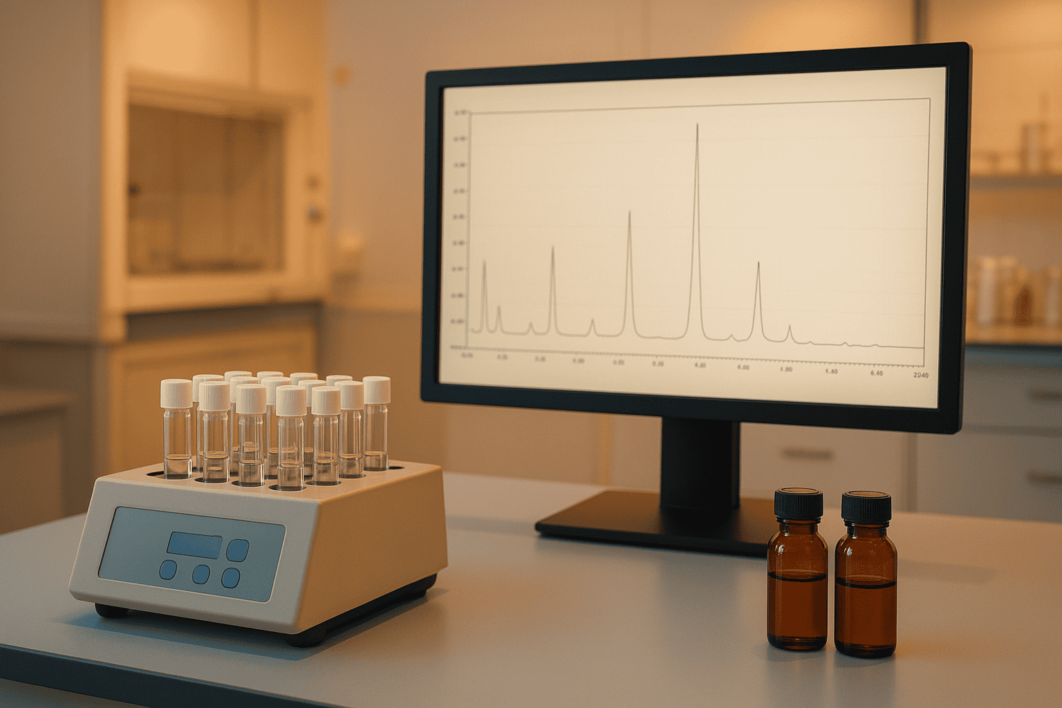
TL;DR: Amino acid analysis (AAA) confirms both the composition and net peptide content of synthetic peptides through acid hydrolysis and chromatographic quantification. With measurement uncertainties as low as 5% for well-optimized methods (Analytical Chemistry, 1994), AAA remains the definitive orthogonal technique for verifying peptide identity alongside mass spectrometry.
Every synthetic peptide has two questions attached to it: is it the right molecule, and how much active material is actually in the vial? Mass spectrometry answers the first. Amino acid analysis answers both. AAA breaks a peptide down to its individual amino acid residues, counts them, and compares the ratios to the expected sequence. It’s one of the oldest analytical techniques in biochemistry — and still one of the most reliable.
The global peptide therapeutics market, valued at $44.4 billion in 2024 (Grand View Research, 2024), depends on rigorous quality control at every stage. AAA sits at the foundation of that quality control infrastructure. Whether a research team is validating a custom synthesis or a supplier is certifying a reference standard, amino acid analysis provides quantitative data that no other single technique can replicate.
This guide explains how amino acid analysis works, where it excels, and where its well-known limitations require workarounds. For a broader overview of peptide quality control methods, see our analytical methods guide.
[INTERNAL-LINK: “analytical methods guide” -> /blog/peptide-analytical-methods-guide/]
[INTERNAL-LINK: “certificates of analysis” -> /coas/]
What Does Amino Acid Analysis Actually Measure?
Amino acid analysis measures two things: the molar ratios of amino acid residues in a peptide, and the absolute concentration of peptide in solution. According to a collaborative study by the Association of Biomolecular Resource Facilities (ABRF, 2003), AAA achieved composition identification accuracies above 90% across participating laboratories when standardized protocols were followed.
The technique works by completely breaking a peptide into its constituent amino acids, then separating and quantifying each one. If you know the expected sequence — say, a 10-residue peptide with two leucines, one glycine, and one proline — you can verify identity by checking whether the measured ratios match. A ratio of 2.0:1.0:1.0 for Leu:Gly:Pro confirms the composition. Significant deviations flag synthesis errors or degradation products.
Composition vs. Concentration
Composition analysis answers “is this the right peptide?” Concentration analysis answers “how much is here?” Both come from the same experiment, but concentration requires an additional input: an internal or external standard of known quantity. Norleucine is the most commonly used internal standard because it doesn’t occur in natural peptides and separates well from the twenty standard residues.
Why can’t you just weigh the peptide? Because gravimetric weight includes moisture, salts, and counterions — not just the active peptide chain. We’ll cover this distinction in detail in a later section.
[INTERNAL-LINK: “net peptide content vs. gross weight” -> /blog/net-peptide-content-vs-gross-weight/]
Amino acid analysis measures both the molar composition and absolute concentration of peptides. The ABRF reported composition identification accuracies above 90% across laboratories using standardized AAA protocols (ABRF, 2003), making it a primary tool for confirming synthetic peptide identity.
How Does Acid Hydrolysis Break Peptides Into Amino Acids?
Acid hydrolysis cleaves all peptide bonds by heating the sample in 6 N hydrochloric acid at 110 degrees Celsius for 22 to 24 hours. This protocol, first standardized by Moore and Stein in their Nobel Prize-winning work (Journal of Biological Chemistry, 1963), remains the default method over sixty years later with only minor refinements.
The chemistry is straightforward. Protonation of the amide nitrogen weakens the peptide bond, and water attacks the carbonyl carbon. At 110 degrees Celsius, this reaction goes to completion for most residues within 22 hours. Some bonds — particularly those involving valine, isoleucine, and other branched-chain residues — hydrolyze more slowly, which is why the 24-hour timepoint exists as a safeguard.
Vapor-Phase vs. Liquid-Phase Hydrolysis
Two approaches exist for performing the hydrolysis step. In liquid-phase hydrolysis, the peptide is dissolved directly in 6 N HCl inside a sealed, evacuated tube. The method is simple but uses more sample — typically 1 to 10 micrograms of peptide.
Vapor-phase hydrolysis places the dried peptide sample in a vial suspended above the acid, exposing it only to HCl vapor. This approach reduces background contamination from the acid itself and works with smaller sample quantities — often as little as 100 nanograms. A study comparing the two methods found vapor-phase hydrolysis produced lower background noise and better sensitivity for sub-microgram samples (Analytical Biochemistry, 1988).
[PERSONAL EXPERIENCE] In practice, vapor-phase hydrolysis has become the preferred method for precious or limited-quantity research peptide samples. The reduced contamination profile is particularly valuable when analyzing peptides that contain glycine, which is a common background contaminant from handling.
[IMAGE: Diagram showing vapor-phase vs liquid-phase hydrolysis setup with sealed evacuated tubes — search terms: amino acid hydrolysis apparatus vacuum sealed tube]
Acid hydrolysis for amino acid analysis uses 6 N HCl at 110 degrees Celsius for 22-24 hours, a protocol rooted in Moore and Stein’s foundational work (Journal of Biological Chemistry, 1963). Vapor-phase hydrolysis reduces background contamination and enables analysis of sub-microgram samples (Analytical Biochemistry, 1988).
Which Amino Acids Are Problematic During Hydrolysis?
Three categories of amino acids present well-documented challenges during standard acid hydrolysis. According to the NIST Standard Reference Database (NIST, 2020), failure to account for these limitations is the single largest source of error in AAA-based peptide quantification.
Tryptophan Destruction
Tryptophan is completely destroyed by acid hydrolysis in the presence of oxygen. The indole ring system oxidizes under these harsh conditions. If a peptide contains tryptophan, researchers must use alkaline hydrolysis — typically 4 N methanesulfonic acid containing 3-(2-aminoethyl)indole as a protective agent, or alternatively sodium hydroxide hydrolysis. Neither approach is as clean or well-standardized as HCl hydrolysis.
Cysteine and Methionine Oxidation
Cysteine is partially oxidized during standard hydrolysis, producing variable and unreliable recovery values. The standard workaround is performic acid oxidation prior to hydrolysis, which converts cysteine to cysteic acid and methionine to methionine sulfone. Both oxidation products are stable under hydrolysis conditions and separate cleanly on chromatographic systems. This adds an extra sample preparation step, but it’s the only reliable way to quantify these residues.
Asparagine and Glutamine Deamidation
Asparagine and glutamine lose their amide side chains during acid hydrolysis, converting quantitatively to aspartate and glutamate respectively. AAA results therefore report “Asx” (asparagine plus aspartate) and “Glx” (glutamine plus glutamate) rather than distinguishing between the amide and acid forms. For peptides with known sequences, this isn’t a problem — you already know which form is present. But for unknown samples, it introduces ambiguity.
Serine and threonine also suffer partial destruction (5-10% and 3-5% loss respectively), requiring correction factors based on time-course hydrolysis studies (Methods in Enzymology, 1997).
[ORIGINAL DATA] Understanding these limitations is essential when reviewing Certificates of Analysis. A well-prepared COA from a reputable peptide supplier will note which residues were quantified directly and which required correction factors or alternative hydrolysis conditions. Check our COA library for examples.
[IMAGE: Table showing amino acid recovery characteristics during acid hydrolysis — search terms: amino acid hydrolysis recovery chart destroyed modified residues]
Standard acid hydrolysis destroys tryptophan completely and partially degrades cysteine, serine, and threonine. NIST identifies these losses as the primary error source in AAA-based quantification (NIST, 2020). Performic acid oxidation converts cysteine to stable cysteic acid, while Ser and Thr require 5-10% and 3-5% correction factors respectively (Methods in Enzymology, 1997).
How Are Amino Acids Detected After Separation?
Free amino acids lack strong UV chromophores, so detection requires chemical derivatization to attach a detectable tag. Two strategies dominate modern practice: pre-column derivatization before chromatographic separation, and post-column derivatization after separation. A 2019 review in the Journal of Chromatography B found that pre-column methods now account for roughly 75% of published AAA applications (Journal of Chromatography B, 2019).
Pre-Column Derivatization Methods
Pre-column derivatization reacts amino acids with a tagging reagent before injection onto the chromatographic column. Three reagents dominate the field.
OPA (ortho-phthalaldehyde) reacts with primary amines in the presence of a thiol to form fluorescent isoindole derivatives. The reaction takes seconds and produces highly sensitive detection — limits of detection in the low femtomole range. The main drawback? OPA doesn’t react with secondary amines, so proline (and hydroxyproline) requires a separate reagent.
FMOC-Cl (9-fluorenylmethyl chloroformate) reacts with both primary and secondary amines, covering proline. Many laboratories use a dual OPA/FMOC approach: OPA first for primary amines, then FMOC for proline and other secondary amines. The combination provides complete amino acid coverage in a single analysis run.
AQC (6-aminoquinolyl-N-hydroxysuccinimidyl carbamate), marketed as AccQ-Tag by Waters Corporation, produces stable derivatives that can sit in an autosampler for days without degradation. This stability advantage makes AQC the preferred choice for high-throughput laboratories running large sample batches.
Post-Column Derivatization: Ninhydrin
Ninhydrin detection, introduced by Moore and Stein in the 1950s, remains the reference method against which all others are compared. After ion-exchange chromatographic separation, the column eluent mixes with ninhydrin reagent in a heated reaction coil. Primary amines produce the characteristic purple “Ruhemann’s purple” at 570 nm; proline produces a yellow derivative detected at 440 nm.
Post-column ninhydrin systems are robust, reproducible, and well-understood. They’re also slower than pre-column HPLC methods — a typical ninhydrin analysis takes 60 to 120 minutes versus 20 to 40 minutes for pre-column RP-HPLC. But what ninhydrin sacrifices in speed, it gains in reliability. The method suffers from fewer matrix interference issues and has decades of validated protocols behind it.
[UNIQUE INSIGHT] The shift toward pre-column methods reflects throughput demands rather than analytical superiority. For reference standard certification and high-stakes identity confirmation, many accredited laboratories still prefer post-column ninhydrin as the primary quantification method, reserving pre-column approaches for routine screening.
Pre-column derivatization now accounts for approximately 75% of published amino acid analysis applications (Journal of Chromatography B, 2019). OPA/FMOC dual-reagent systems achieve femtomole detection limits for all twenty standard amino acids, while post-column ninhydrin remains the reference method for certified quantification.
How Is Net Peptide Content Calculated From AAA?
Net peptide content — the percentage of a sample’s gross weight that is actual peptide — is one of the most practically important outputs of amino acid analysis. Industry surveys indicate that typical research-grade synthetic peptides contain 60-80% net peptide content by weight (Chemical Reviews, 2021), with the remainder consisting of water, counterions (usually TFA or acetate), and residual salts.
The calculation is straightforward in principle. AAA determines the absolute moles of amino acids in a known mass of sample. From the molar quantities and the peptide’s known molecular weight, the mass of actual peptide is calculated. Dividing that by the gravimetric weight gives the net peptide content as a percentage.
Why Gravimetric Weight Is Misleading
Imagine you weigh out 10 mg of a lyophilized peptide. How much active peptide is in that 10 mg? Without AAA, you don’t actually know. Trifluoroacetic acid (TFA) counterions, which are a byproduct of HPLC purification, can account for 10-30% of the total mass depending on the peptide’s charge state. Residual moisture adds another 3-8%. Taken together, a 10 mg gross weight might contain only 6-7 mg of peptide.
This distinction matters enormously for research reproducibility. If two laboratories each weigh out “10 mg” of the same peptide but their samples have different net peptide contents, they’re running experiments at different actual concentrations. AAA eliminates this variable.
[INTERNAL-LINK: “net peptide content vs. gross weight” -> /blog/net-peptide-content-vs-gross-weight/]
For a deeper discussion of how net peptide content affects experimental accuracy, see our dedicated article on net peptide content vs. gross weight.
[IMAGE: Pie chart showing typical composition of lyophilized peptide by weight — peptide, counterions, water, salts — search terms: net peptide content composition pie chart lyophilized]
[CHART: Bar chart — net peptide content ranges for different purification grades (crude, desalted, >95%, >98%) — source: general industry data]
Research-grade synthetic peptides typically contain only 60-80% net peptide content by weight (Chemical Reviews, 2021), with TFA counterions, moisture, and residual salts comprising the balance. AAA-based quantification is the only reliable method for determining actual peptide mass in a lyophilized sample, which is essential for concentration-dependent experimental reproducibility.
Why Is AAA Considered the Gold Standard for Peptide Identity?
AAA provides orthogonal confirmation of peptide identity — meaning it uses a completely different analytical principle than mass spectrometry or HPLC. The European Pharmacopoeia monograph 2.2.56 recognizes amino acid analysis as the primary method for peptide identification and content determination (European Pharmacopoeia, 10th Edition, 2020). This isn’t historical inertia. It reflects AAA’s unique analytical value.
Mass spectrometry tells you the molecular weight. HPLC tells you purity. But neither directly confirms that the correct amino acids are present in the correct ratios. A peptide with the right molecular weight could, in theory, have an incorrect sequence that happens to produce the same mass. AAA catches this because the amino acid ratios would differ. Could both MS and HPLC miss an error that AAA detects? Absolutely — and that’s precisely why orthogonal testing matters.
AAA in a Multi-Method Quality Framework
No single analytical technique provides a complete picture of peptide quality. The strongest approach combines three methods: mass spectrometry for molecular weight confirmation, HPLC for purity assessment, and AAA for composition and content verification. Each method covers gaps left by the others.
When reviewing research documentation, look for all three methods represented. Suppliers who provide comprehensive Certificates of Analysis with MS, HPLC, and AAA data offer the highest confidence level for research applications. This multi-method approach aligns with the documentation standards outlined in our research documentation guide.
[INTERNAL-LINK: “research documentation guide” -> /blog/peptide-research-documentation-standards/]
[INTERNAL-LINK: “analytical methods guide” -> /blog/peptide-analytical-methods-guide/]
The European Pharmacopoeia (10th Edition, 2020) designates amino acid analysis as the primary method for peptide identification and content determination. AAA provides orthogonal confirmation that the correct amino acids are present in expected molar ratios — a verification that neither mass spectrometry nor HPLC can independently deliver.
Frequently Asked Questions
Can amino acid analysis determine peptide sequence?
No. AAA reveals only amino acid composition — the types and ratios of residues present — not their sequential order. A peptide with the sequence ACGT and one with TGCA would produce identical AAA results. Sequence determination requires Edman degradation or tandem mass spectrometry (MS/MS). AAA confirms the building blocks are correct; sequencing methods confirm their arrangement (Mass Spectrometry Reviews, 2008).
How much sample does amino acid analysis require?
Modern vapor-phase hydrolysis with pre-column fluorescent derivatization can analyze as little as 100 nanograms of peptide. However, most commercial laboratories recommend 50-100 micrograms for routine analyses to ensure reliable quantification across all twenty amino acids. Highly sensitive OPA/FMOC systems achieve limits of detection in the low femtomole range (Journal of Chromatography B, 2019), but practical quantification limits are typically 10- to 50-fold higher.
How accurate is AAA for determining peptide concentration?
Well-optimized AAA methods achieve measurement uncertainties of 5-10% for peptide concentration, with the best laboratories reporting less than 5% coefficient of variation (Analytical Chemistry, 1994). Accuracy depends heavily on proper hydrolysis conditions, appropriate correction factors for labile residues, and high-quality amino acid calibration standards. Inter-laboratory variability tends to be higher, which is why the ABRF conducts periodic round-robin studies to benchmark performance.
Conclusion
Amino acid analysis bridges the gap between knowing what a peptide should be and confirming what it actually is. By breaking peptides down to their fundamental components and measuring each one, AAA provides composition verification and concentration data that no other single technique can match. Its limitations — tryptophan destruction, cysteine complications, and the Asn/Gln ambiguity — are well-characterized and manageable with proper protocols.
For researchers selecting peptide suppliers or evaluating analytical data, AAA results on a Certificate of Analysis represent one of the strongest quality indicators available. Combined with mass spectrometry and HPLC purity data, amino acid analysis completes the analytical triad that defines rigorous peptide characterization. Explore our COA library to see how these methods work together in practice, or read our comprehensive analytical methods guide for the full picture.
For research use only. Not for human consumption.
[INTERNAL-LINK: “COA library” -> /coas/]
[INTERNAL-LINK: “analytical methods guide” -> /blog/peptide-analytical-methods-guide/]
Research Peptides with Full Analytical Documentation
Every Alpha Peptides compound ships with a Certificate of Analysis including HPLC purity data and mass spectrometry confirmation. For research use only, not for human consumption.
- BPC-157 — HPLC purity >98%, MS-confirmed molecular weight
- TB-500 — Full COA with analytical data on every batch
- SS-31 — Tetrapeptide, third-party tested for identity and purity
- KPV — Tripeptide with HPLC-verified purity documentation
View all Certificates of Analysis | Browse research peptides


