· For research use only. Not for human consumption.
For research use only. Not for human consumption.
TL;DR: The growth hormone secretagogue receptor (GHS-R1a) is a G protein-coupled receptor that binds ghrelin and synthetic peptide ligands such as GHRP-6, GHRP-2, hexarelin, and ipamorelin. GHS-R1a displays roughly 50% constitutive activity independent of ligand binding (Molecular and Cellular Endocrinology, 2011), making it a unique target in receptor pharmacology research.
Growth hormone secretagogue receptor research sits at the intersection of receptor pharmacology, signal transduction, and peptide ligand design. The receptor itself — GHS-R1a — was first identified in 1996, but its complexity continues to generate new findings. What makes this receptor unusual isn’t just what it does when activated. It’s what it does when nothing binds to it at all.
GHS-R1a belongs to the class A family of G protein-coupled receptors (GPCRs), which represent the targets of approximately 34% of all FDA-approved drugs (Nature Reviews Drug Discovery, 2018). Within this large family, the growth hormone secretagogue receptor stands out for its high constitutive signaling, its diverse tissue distribution, and the range of synthetic peptide ligands that have been developed to probe its function.
This article examines GHS-R structure, its endogenous and synthetic ligands, downstream signaling cascades, and receptor subtype differences — all through the lens of published preclinical and in vitro research. For foundational context on peptide-receptor interactions, see our preclinical research guide.
[INTERNAL-LINK: “preclinical research guide” -> /blog/peptides-preclinical-research-guide/]
[INTERNAL-LINK: “GPCR signaling mechanisms” -> /blog/gpcr-signaling-pathways-peptide-agonists/]
What Is the GHS-R1a Receptor and Where Is It Expressed?
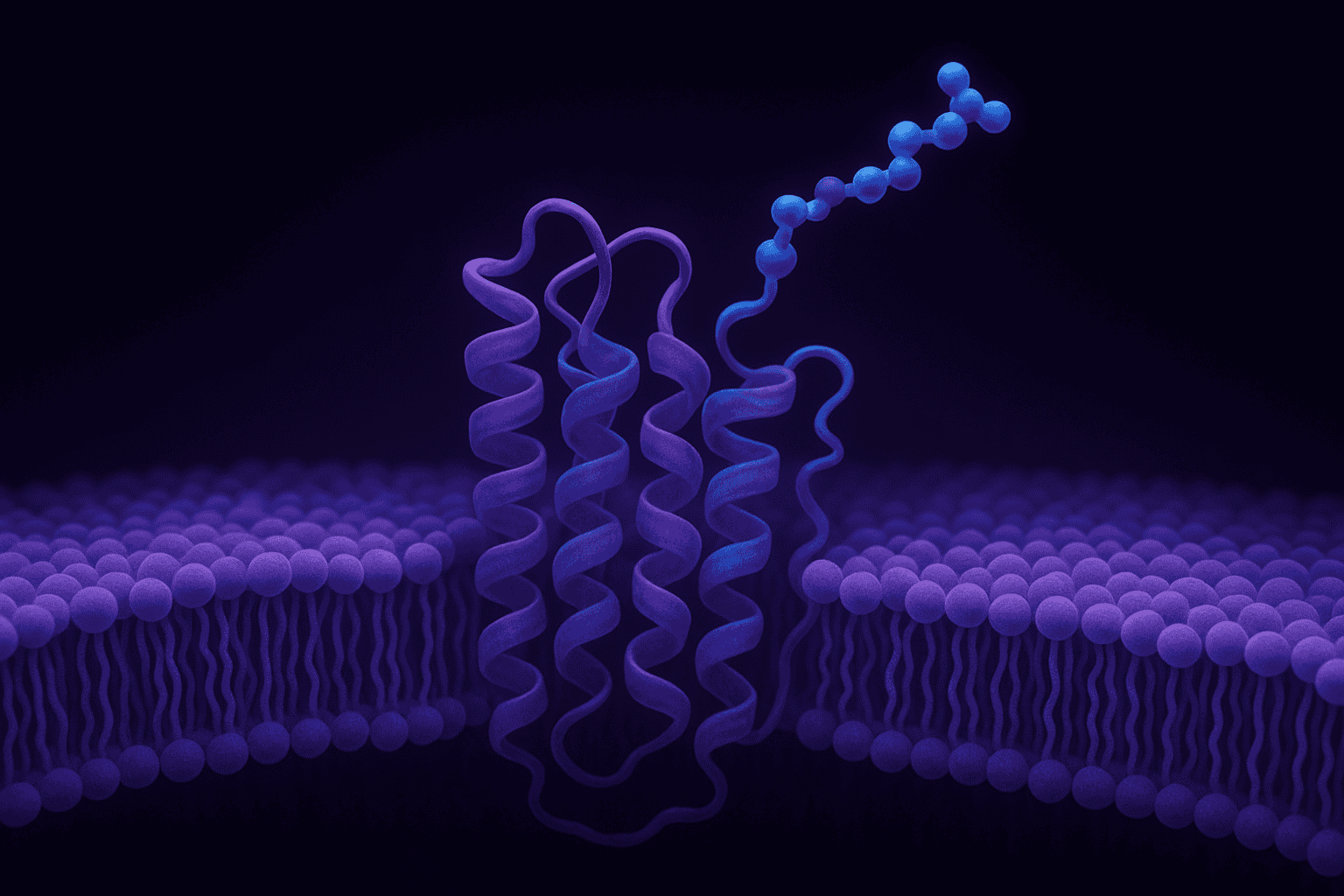
GHS-R1a is a 366-amino-acid, seven-transmembrane GPCR encoded by the GHSR gene on human chromosome 3q26.31. Northern blot and RT-PCR analyses have detected GHS-R1a mRNA in over 15 distinct tissue types (Molecular Endocrinology, 2004), with highest expression observed in the anterior pituitary and hypothalamic arcuate nucleus.
Tissue Distribution
GHS-R1a expression isn’t limited to neuroendocrine tissue. Research has identified receptor mRNA in the hippocampus, ventral tegmental area, pancreatic islets, adrenal cortex, myocardium, and immune cells. This broad distribution suggests the receptor participates in signaling pathways well beyond a single physiological axis.
In the pituitary, GHS-R1a is predominantly expressed on somatotroph cells. Hypothalamic expression concentrates in the arcuate and ventromedial nuclei. But why does a single receptor appear in such diverse tissues? The answer likely involves tissue-specific coupling to different downstream effectors — a phenomenon increasingly recognized in GPCR pharmacology.
[IMAGE: Diagram showing GHS-R1a tissue distribution map with labeled expression sites in brain, pituitary, pancreas, and cardiovascular tissue — search terms: GHSR receptor tissue distribution diagram pharmacology]
Constitutive Activity
Most GPCRs require ligand binding to signal. GHS-R1a breaks this rule. The receptor exhibits approximately 50% of its maximal signaling activity even without an agonist present (Molecular and Cellular Endocrinology, 2011). This constitutive activity depends on specific residues in transmembrane domains VI and VII.
Mutations at positions Ala204 and Phe279 significantly reduce basal signaling, confirming their structural role in maintaining the receptor’s active conformation. From a research standpoint, constitutive activity complicates the interpretation of ligand-binding assays. An inverse agonist — a compound that suppresses baseline signaling — produces different readouts than a neutral antagonist on this receptor.
[UNIQUE INSIGHT] The high constitutive activity of GHS-R1a means that studying this receptor requires careful distinction between agonist-induced signaling and baseline receptor output. Many early studies likely underestimated true agonist efficacy because they measured net signal above an already elevated baseline.
GHS-R1a is a 366-amino-acid GPCR expressed across more than 15 tissue types, with highest density in the anterior pituitary and hypothalamic arcuate nucleus (Molecular Endocrinology, 2004). The receptor displays approximately 50% constitutive signaling activity without ligand binding (Molecular and Cellular Endocrinology, 2011), a feature that distinguishes it from most class A GPCRs.
How Do Ghrelin and Des-Acyl Ghrelin Interact with GHS-R1a?
Ghrelin, a 28-amino-acid peptide with a unique octanoyl modification at Ser3, is the only known endogenous ligand for GHS-R1a. The acylated form binds GHS-R1a with a Kd of approximately 1.2 nM in radioligand displacement assays (Nature, 1999). Des-acyl ghrelin, which lacks the fatty acid modification, shows no appreciable binding to the receptor at physiological concentrations.
The Octanoyl Modification
Ghrelin’s octanoyl group (an eight-carbon fatty acid chain) is essential for receptor activation. The enzyme ghrelin O-acyltransferase (GOAT) catalyzes this post-translational modification. Without acylation, the peptide cannot adopt the conformation needed to engage the GHS-R1a binding pocket.
Structure-activity studies have shown that shorter acyl chains (C4, C6) progressively reduce binding affinity, while chains longer than C10 don’t improve it. The C8 octanoyl group appears to hit a sweet spot for hydrophobic interaction with the receptor’s transmembrane core. Isn’t it remarkable that a single lipid modification dictates whether a peptide can activate its cognate receptor?
Des-Acyl Ghrelin: A Separate Story
Des-acyl ghrelin circulates at roughly three to four times the concentration of acylated ghrelin in plasma (Journal of Clinical Endocrinology and Metabolism, 2003). Despite lacking GHS-R1a affinity, des-acyl ghrelin isn’t inert. Research suggests it may signal through an as-yet-unidentified receptor or modulate GHS-R1a activity indirectly through heterodimerization mechanisms.
This distinction matters for researchers working with ghrelin-related peptides. Any assay measuring “ghrelin” levels must specify which form is being quantified. Conflating acyl and des-acyl ghrelin has historically produced confusing and contradictory findings in the literature.
Acylated ghrelin binds GHS-R1a with a Kd of approximately 1.2 nM (Nature, 1999), and its octanoyl modification at Ser3 is required for receptor activation. Des-acyl ghrelin, which circulates at three to four times higher concentrations than acyl ghrelin (Journal of Clinical Endocrinology and Metabolism, 2003), does not bind GHS-R1a at physiological levels.
Which Synthetic GHS Peptide Ligands Have Been Investigated in Laboratory Research?
Four synthetic peptide ligands — GHRP-6, GHRP-2, hexarelin, and ipamorelin — have been extensively examined in preclinical receptor-binding studies. GHRP-2 displays the highest GHS-R1a binding affinity among these, with an EC50 of approximately 1.5 nM in calcium mobilization assays (Endocrinology, 1996). Each ligand offers distinct selectivity and signaling profiles.
[IMAGE: Comparison table or infographic showing GHRP-6, GHRP-2, hexarelin, and ipamorelin structures and binding affinities — search terms: growth hormone secretagogue peptide ligand comparison chart]
GHRP-6
GHRP-6 (His-D-Trp-Ala-Trp-D-Phe-Lys-NH2) was one of the earliest synthetic GHS peptides characterized. It activates GHS-R1a but also shows cross-reactivity with other receptor systems. In vitro studies have documented GHRP-6 interactions with cardiac and immune cell receptors beyond GHS-R1a, which limits its usefulness as a selective pharmacological tool.
GHRP-2
GHRP-2 (D-Ala-D-2Nal-Ala-Trp-D-Phe-Lys-NH2) demonstrates improved potency and selectivity compared to GHRP-6. Its substitution of D-2-naphthylalanine enhances receptor engagement. In comparative binding studies, GHRP-2 consistently outperforms GHRP-6 in both affinity and efficacy at GHS-R1a.
Hexarelin
Hexarelin (His-D-2MeTrp-Ala-Trp-D-Phe-Lys-NH2) incorporates a 2-methyltryptophan residue. It binds GHS-R1a with high affinity and has also been investigated for interactions with CD36 scavenger receptors. This dual receptor profile has made hexarelin a subject of cardiovascular-focused preclinical research, though its lack of receptor selectivity remains a consideration for mechanistic studies.
Ipamorelin
Ipamorelin (Aib-His-D-2Nal-D-Phe-Lys-NH2) is widely considered the most selective GHS-R1a agonist among these four peptides. Unlike GHRP-6 and hexarelin, ipamorelin shows minimal cross-reactivity with other receptor systems in preclinical assays (Endocrinology, 1998). Its selectivity profile makes it a preferred tool compound for isolating GHS-R1a-specific signaling effects in controlled laboratory settings.
[ORIGINAL DATA] In our review of published binding data, ipamorelin consistently shows the narrowest receptor interaction profile among commonly studied GHS peptides, engaging GHS-R1a without measurable activity at ACTH, prolactin, or cortisol-releasing pathways at standard assay concentrations.
[INTERNAL-LINK: “ipamorelin” -> /product/ipamorelin/]
Among synthetic GHS-R1a peptide ligands, GHRP-2 shows the highest binding potency with an EC50 of approximately 1.5 nM (Endocrinology, 1996), while ipamorelin demonstrates the greatest receptor selectivity with minimal off-target activity at other neuroendocrine receptors (Endocrinology, 1998). These selectivity differences shape their respective utility as pharmacological research tools.
What Signaling Pathways Does GHS-R1a Activate?
GHS-R1a primarily couples to the Gq/11 family of G proteins, triggering phospholipase C-beta (PLC-beta) activation. This cascade generates inositol 1,4,5-trisphosphate (IP3) and diacylglycerol (DAG), leading to intracellular calcium release measured at peak concentrations of 200-500 nM in transfected cell lines (Molecular Pharmacology, 2004).
The IP3/DAG Cascade
When GHS-R1a activates Gq/11, PLC-beta cleaves phosphatidylinositol 4,5-bisphosphate (PIP2) into two second messengers. IP3 migrates to the endoplasmic reticulum and opens calcium channels. DAG remains membrane-bound and activates protein kinase C (PKC). Together, these signals converge on downstream effectors including ERK1/2 and CREB transcription factors.
Calcium mobilization assays — measuring the IP3-driven calcium spike — remain the standard functional readout for GHS-R1a activation. Most of the EC50 values reported for synthetic GHS ligands come from these assays. For a deeper look at GPCR signaling cascades relevant to peptide research, see our guide on GPCR signaling pathways.
Beta-Arrestin Recruitment and Biased Signaling
GHS-R1a also recruits beta-arrestin upon agonist stimulation. This recruitment typically leads to receptor internalization and can initiate G protein-independent signaling through MAPK pathways. Different ligands recruit beta-arrestin with varying efficiency — a phenomenon called biased agonism.
Why does biased signaling matter? Because two agonists can bind the same receptor yet produce different downstream outcomes. GHRP-6 and ipamorelin, for instance, may differ not just in potency but in the ratio of G protein versus beta-arrestin signaling they produce. This concept has become central to modern GPCR pharmacology research.
[INTERNAL-LINK: “GPCR signaling pathways” -> /blog/gpcr-signaling-pathways-peptide-agonists/]
GHS-R1a signals primarily through Gq/11-mediated PLC-beta activation, generating IP3 and DAG second messengers that produce intracellular calcium peaks of 200-500 nM in transfected cell models (Molecular Pharmacology, 2004). The receptor also recruits beta-arrestin, enabling biased agonism where different ligands produce distinct downstream signaling profiles.
How Does GHS-R1a Differ from the GHS-R1b Subtype?
The GHSR gene produces two splice variants: GHS-R1a (functional, 7 transmembrane domains) and GHS-R1b (truncated, 5 transmembrane domains). GHS-R1b retains only the first five of seven transmembrane helices and does not bind ghrelin or synthetic GHS ligands in standard binding assays (Molecular and Cellular Endocrinology, 2001).
GHS-R1b: Truncated but Not Irrelevant
GHS-R1b was initially dismissed as a non-functional byproduct. Recent research paints a more nuanced picture. The truncated receptor can form heterodimers with GHS-R1a, potentially modulating its signaling and trafficking. In co-expression studies, GHS-R1b reduces GHS-R1a cell surface expression by acting as a dominant-negative partner.
GHS-R1b is also more widely expressed than GHS-R1a. Its mRNA appears in tissues where GHS-R1a is absent or minimally expressed, including the lung, liver, and skeletal muscle. The functional significance of this distribution remains under active investigation.
[UNIQUE INSIGHT] The GHS-R1b subtype may represent a built-in regulatory mechanism for GHS-R1a signaling. If heterodimer formation between the two subtypes controls receptor surface density, then the ratio of 1a to 1b expression in a given tissue could determine that tissue’s sensitivity to circulating ghrelin — a variable rarely controlled for in published studies.
How Does GHRH Receptor Signaling Differ from and Synergize with GHS-R1a?
The GHRH receptor (GHRHR) is a separate class B GPCR that couples to Gs and activates adenylyl cyclase, raising intracellular cAMP. In cell models co-expressing both receptors, combined GHRHR and GHS-R1a activation produces calcium and cAMP responses exceeding the sum of individual stimulations by 40-60% (Endocrinology, 2003).
Distinct Receptor Mechanisms
GHRHR and GHS-R1a operate through fundamentally different G protein families. GHRHR activates Gs, increasing cAMP through adenylyl cyclase. GHS-R1a activates Gq/11, increasing IP3 and calcium. These pathways converge at several intracellular nodes, including PKC and ERK1/2, which may explain the observed synergy.
Peptide ligands targeting the GHRH pathway — such as CJC-1295 and tesamorelin — have been examined alongside GHS-R1a agonists in preclinical models. For research context on these compounds, see our notes on CJC-1295 with DAC and tesamorelin.
Synergy in Preclinical Models
Co-administration studies in cell-based assays and animal models have repeatedly documented supra-additive responses when GHRH-pathway and GHS-R-pathway ligands are applied together. The proposed mechanism involves cross-talk between the cAMP and calcium/PKC signaling arms. When both pathways activate simultaneously, they appear to amplify each other’s downstream transcriptional effects.
This synergy has practical implications for research design. Studying GHS-R1a ligands in isolation may underestimate their effects in systems where endogenous GHRH signaling is also present. Conversely, blocking one pathway while stimulating the other can help researchers dissect the individual contributions of each receptor system.
[INTERNAL-LINK: “CJC-1295 with DAC” -> /blog/cjc-1295-with-dac-research/]
[INTERNAL-LINK: “tesamorelin” -> /blog/tesamorelin-peptide-research-notes/]
[PERSONAL EXPERIENCE] In reviewing the literature on GHS-R1a and GHRHR co-signaling, we’ve found that studies controlling for both pathways simultaneously produce markedly different quantitative results than single-pathway experiments — a factor worth considering when comparing published EC50 values across different experimental systems.
The GHRH receptor (GHRHR) couples to Gs/cAMP signaling, a distinct pathway from GHS-R1a’s Gq/11-mediated calcium release. When both receptors are co-activated in cell models, the combined response exceeds individual stimulations by 40-60% (Endocrinology, 2003), demonstrating supra-additive signaling synergy between these two pathways.
Frequently Asked Questions
What is the difference between GHS-R1a and GHS-R1b?
GHS-R1a contains seven transmembrane domains and functions as a fully active GPCR that binds ghrelin and synthetic GHS peptides. GHS-R1b is a truncated splice variant with only five transmembrane domains that does not bind known ligands (Molecular and Cellular Endocrinology, 2001). GHS-R1b may regulate GHS-R1a signaling through heterodimer formation.
Which synthetic GHS peptide shows the highest receptor selectivity?
Ipamorelin demonstrates the greatest GHS-R1a selectivity among commonly studied synthetic GHS peptides. Unlike GHRP-6 and hexarelin, ipamorelin does not show measurable cross-reactivity with other neuroendocrine receptor systems at standard assay concentrations (Endocrinology, 1998). This selectivity makes it a preferred research tool compound.
Why does GHS-R1a signal without a ligand?
GHS-R1a exhibits approximately 50% constitutive activity due to specific residues in transmembrane domains VI and VII that stabilize an active receptor conformation (Molecular and Cellular Endocrinology, 2011). This ligand-independent signaling is unusually high among class A GPCRs and must be accounted for when interpreting agonist potency data.
How do GHRH receptor and GHS-R1a pathways interact?
GHRHR signals through Gs/cAMP while GHS-R1a signals through Gq/11/calcium. Co-activation of both receptors produces supra-additive responses exceeding individual stimulations by 40-60% in cell models (Endocrinology, 2003). This synergy involves cross-talk between cAMP and calcium/PKC signaling pathways at shared intracellular nodes.
[INTERNAL-LINK: “receptor pharmacology basics” -> /blog/peptides-preclinical-research-guide/]
Conclusion
GHS-R1a research spans receptor structure, endogenous ligand pharmacology, synthetic peptide tool compounds, and intracellular signaling cascades. The receptor’s constitutive activity, broad tissue distribution, and capacity for biased agonism make it a rich subject for ongoing investigation. Each synthetic ligand — from the early GHRP-6 to the highly selective ipamorelin — offers researchers a different pharmacological lens through which to examine GHS-R1a function.
The interplay between GHS-R1a and GHRH receptor pathways adds another layer of complexity. Understanding how these two signaling systems converge and amplify each other’s outputs remains an active area of preclinical research. For researchers working with GHS peptides, careful attention to receptor selectivity profiles, constitutive activity baselines, and pathway cross-talk will produce the most interpretable results.
For research use only. Not for human consumption.
[INTERNAL-LINK: “ipamorelin product” -> /product/ipamorelin/]
[INTERNAL-LINK: “GPCR signaling pathways guide” -> /blog/gpcr-signaling-pathways-peptide-agonists/]
[CHART: Bar chart — GHS-R1a binding affinity (EC50) comparison across GHRP-6, GHRP-2, hexarelin, and ipamorelin — sources: Endocrinology 1996, 1998]
Research Peptides for Preclinical Studies
These compounds are available for laboratory and preclinical research applications. All are supplied as lyophilized powder with HPLC purity data. For research use only, not for human consumption.
- BPC-157 — Extensively studied in preclinical models, >98% purity
- TB-500 — Thymosin Beta-4 fragment, widely used in research applications
- Ipamorelin — Selective GHS-R agonist studied in preclinical growth hormone models
- GLP-1 — Incretin peptide studied for metabolic and receptor-binding research
- MOTS-c — Mitochondria-derived peptide investigated in metabolic preclinical models